How financial conflicts of interest affect medicine

Ahn R, et al. Financial ties of principal investigators and randomized controlled trial outcomes: cross sectional study. BMJ. 2017;356.

Not neonatology but a study of how principal investigators’ financial ties to the pharmaceutical industry are associated with outcomes of controlled trials of drug efficacy funded by either industry drug wholesalers or non-industry sources. The authors searched Google with the name of the PI and the drug company producing the medication being tested in a randomized controlled trial, they also used a couple of databases to identify the financial links between investigators and drug companies. Two thirds of the PIs had some financial ties with the companies producing the drugs that they were testing.

Of all principal investigators, 156 (39%) reported advisor/consultancy payments, 81 (20%) reported speakers’ fees, 81 (20%) reported unspecified financial ties, 52 (13%) reported honorariums, 52 (13%) reported employee relationships, 52 (13%) reported travel fees, 41 (10%) reported stock ownership, and 20 (5%) reported having a patent related to the study drug. The prevalence of financial ties of principal investigators was 76% (103/136) among positive studies and 49% (29/59) among negative studies. In unadjusted analyses, the presence of a financial tie was associated with a positive study outcome (odds ratio 3.23, 95% confidence interval 1.7 to 6.1). In the primary multivariate analysis, a financial tie was significantly associated with positive RCT outcome after adjustment for the study funding source (odds ratio 3.57 (1.7 to 7.7).

Most of the financial ties were not clearly reported on the primary publication. Even for publicly funded trials financial links with industry are associated with a much higher chance that you will show that the drug that you have a vested financial interest in is effective. I don’t know what we can do about such things, realistically. I have received travel expenses (usually indirectly through the organizing committee of a conference) from industry (specifically from Mallickrodt and from Chiesi) . I think we should make more effort to be transparent about all of these links, and more effort to reduce them and make them more arms length also.

Two other articles address related issues this week, the first (Lin DH, et al. Financial conflicts of interest and the centers for disease control and prevention’s 2016 guideline for prescribing opioids for chronic pain. JAMA internal medicine. 2017) examined comments sent to the CDC following release of a new guideline which aimed to restrict opioid prescribing in the hope of having an impact on the current epidemic of opioid related deaths in the USA (which is spreading across Canada also, from Vancouver in our direction). There were 158 organisations who submitted comments about the draft guidelines, one third of them received financial support from opioid manufacturers, those organisations were much more likely to be negative about the guideline, (although many were positive and some submitted suggestions to improve). Organisations were not required to state any potential conflicts when submitting comments so these authors also had to do a bit of detective work to find these links.

The CDC should require commenters to reveal their potential conflicts of interest.

Another article examined financial conflicts of interest among Hematologist-Oncologists who use Twitter, although we don’t know what they were tweeting about in this study, other research has shown that physicians using twitter tweet about medical subjects more than 60% of the time and often mention drugs or medical products. They found that 80% of the Twitter active Hematologists had a financial conflict of interest. I don’t think that there are many who report, or have a link to a report of, their potential conflicts of interest.

Twitterers beware!


Posted in Not neonatology | Leave a comment

Happy New Review no. 2; Inhaled NO in the full term infant

We have also updated the Cochrane review of inhaled NO in the term infant. (Barrington KJ, Finer N, Pennaforte T, Altit G. Nitric oxide for respiratory failure in infants born at or near term. Cochrane database of systematic reviews (Online). 2017;1:CD000399). This was also a major update, with the addition of 3 trials, but also a restructuring of the review. On adding the trial from Gonzalez, which was a trial of early compared to late NO in 2 hospitals in Chile without access to ECMO, we re-thought several of the other trials that we had previously analyzed as being trials of NO which allowed backup treatment of the controls with NO. In fact there were 6 trials in total which had criteria for enrolment, and randomized babies to immediate inhaled NO, or to a control group who would receive inhaled NO if their oxygenation worsened to more severe criteria.

We have now analyzed those 6 trials (Barefield 1996; Day 1996; Sadiq 1998; Cornfield 1999; Konduri 2004; Gonzalez 2010) as comparisons of early compared to late intervention with NO.

The risk of bias figure, including all the trials, is below:

risk-of-bias-summary

For several of the trials the funding source was not clear, hence the  question mark, but for several which are marked by the question mark in yellow, including for example Konduri 2004, funding was primarily from a government granting agency, but there was also partial support from industry, with the manufacturer of inhaled NO providing the gas (and placebo) free of charge for the trial.

For the first comparison, inhaled NO versus control, studies which did not allow back-up treatment with NO in controls, the results of the analyses are unchanged. Inhaled NO is clearly beneficial in terms of reducing the need for ECMO, with an NNT of about 5.

We now present in the review a second comparison, inhaled NO at moderate compared to severe criteria for illness severity. The Summary of Findings table looks like this:

no-in-the-term

The actual criteria for illness severity were not consistent between trials, moderate severity was either based on Oxygenation Index (between 15 and 25, or 25 to 35 or 10 to 30) or on AaDO2 of 500 to 599. Whereas severe criteria were if they passed the upper limit of those ranges; so greater than 25 or 35 or 30 or an AaDO2 of 600 or more. I think there is enough overlap in the qualifying criteria that the studies satisfy Barrington’s rule, and that it is reasonable to meta-analyze them. But I realize that others may have a different response to that.

The overall message is that there is no proven advantage in clinical outcomes (death or needing ECMO) of starting NO earlier, compared to waiting for more severe criteria of illness severity. Fewer infants will progress to severe criteria if you start NO early, but if you wait until they become more hypoxic they will still respond.

There are, as always, some limitations to that statement, the confidence intervals are very wide, and include a possibility of a substantial relative reduction in mortality, (or a large increase). The absolute numbers dying, for example are 10% in the group who were treated at severe illness criteria, this might change to between 3.8% and 12.6% if the babies are treated earlier.

I don’t think based on these numbers that any promotion of the earlier use of inhaled NO can be justified. You will treat 100% of babies at moderate illness criteria, compared to treating about 60% of them if you wait for more severe criteria, which will have an impact on resource utilisation, but no evident impact on clinically important outcomes.

Posted in Neonatal Research | Tagged , , | Leave a comment

Happy New Year! And Happy Reading; an update of the Cochrane review: nitric oxide in the preterm

A major revision of the Cochrane review of inhaled nitric oxide gas in the preterm infant is now available. (Barrington KJ, Finer N, Pennaforte T. Inhaled nitric oxide for respiratory failure in preterm infants. Cochrane database of systematic reviews (Online). 2017;1:CD000509). It includes 2 new publications, a small RCT from China, and the large multicenter RCT in preterm babies still requiring respiratory support at 5 to 14 days: the NEWNO trial (listed in the review as Yoder 2013). This was an unusual situation for me as the NEWNO trial has still not been published; we waited a while to finalize the review as we thought that it would be better that NEWNO was in print before including it in the meta-analysis. Eventually, because we had access to much of the summary data, we decided, with the delays in publication of the primary data for the entire study, that we should proceed. It is possible of course that we may need to make some minor adjustments to the review once the peer-reviewed publication appears, but it is unlikely, I think, that there will be any major changes to make. The main conclusions are very likely to stand.

One of the great things about Cochrane reviews is that they are (supposed to be) kept up to date. With frequent new literature searches and inclusion of new trials as they appear. Which means that authoring a review is a serious commitment. Also this time for the new reviews there are a number of innovations in the Cochrane Collaboration standards since the last time I updated the review. As well as the graphics to show the risk of bias evaluations, each new review (or updated review) will have a Summary of Findings Table, using Grade standards (and the Grade groups on-line software).

In case you haven’t seen them before here is one of the risk of bias figures: it should be clear that this is really what is meant, not that there has been bias, but that a lack of allocation concealment (for example) increases the risk of bias. For this review, many of the trials were funded by industry (previously not a common occurrence in neonatology) so we decided to add the column entitled ‘Funding Source’. There are abundant data to show that industry funding skews the medical literature, I therefore think we should have such a column for all Cochrane reviews, but we must be clear that although this increases the risk of bias, it doesn’t necessarily mean that those trials marked in red were actually biased. By most standards, in fact, the industry-funded trials in this review appear to have been free of the problems with many such trials, like inappropriate composite short-term outcomes, controls groups almost designed to have poorer outcomes, selective publication and selective reporting of outcomes.

cochrane-premie-no

The Summary of Findings tables are newer, and follow GRADE standards for determining how much confidence we have in the findings. This is the SoF table for the new version of this review.

no-preterm-sof-table

As you can see, the summary is that there is no evidence of benefit from the use of inhaled NO for any of these indications in the preterm. The situation where the potential benefit is closest to being statistically significant is the use of inhaled NO for infants older than 3 days who are determined to be at elevated risk of BPD. One of those studies was somewhat different to the other 2 in that category, the study by Subhedar, but that trial only contributed about 40 of the over 1000 subjects, so we did a sensitivity analysis, eliminating that study, which still shows an effect consistent with chance.

no-preterm-forest-plot

I also applied the Barrington rule, which is that if it was not possible to be eligible for 2 studies, they shouldn’t be meta-analyzed, Subhedar’s study included babies at 96 hours, the other two studies were minimum 5 days of age, and those two had considerable overlap in eligibility. So the 2 larger studies satisfy the Barrington rule, but not the smaller one.

So is this the final word? Does this mean that it is never justified to give inhaled NO to a preterm infant? My response to my own question is that I think that the evidence is consistent with a statement such as “the data currently show that the administration of inhaled NO to critically ill preterm infants in the first few days of life, with hypoxic respiratory failure, does not lead to an overall increase in survival”. That is not the same as saying that there are never any indications for a trial of inhaled NO, especially when there are clear indications of pulmonary hypertension.

Some groups of preterm babies have not been adequately studied, for example, preterm infants with clear evidence of pulmonary hypertension. Such infants may have a poor response to surfactant, and have a severe pre- to post- ductal oxygenation gradient, they often have a dramatic immediate response to inhaled NO, just like babies at term. It seems unreasonable to withhold NO from such a baby at 33 weeks gestation, when an otherwise identical baby at 35 weeks gestation would receive the treatment. Our individual patient data meta-analysis suggested that there may possibly be a benefit to using inhaled NO in preterm babies with pulmonary hypertension, but the interaction term was not statistically significant, and there were very few babies for whom pulmonary hypertension had been recorded in their original data files, and there was not a clear, consistent definition of pulmonary hypertension either.

I do think there is room for more trials in the preterm. An ethically justifiable trial would include preterm babies with good evidence of pulmonary hypertension, and would allow rescue treatment in the controls if they continued to be hypoxic after an interval, such as 60 minutes. Current data show a high frequency of intracranial hemorrhage in preterm babies with apparent pulmonary hypertension who receive NO; and I think it is possible that the sudden improvement in pulmonary vascular resistance, and sudden increase in cerebral perfusion that accompanies this change, could cause an increase in IVH. It might be that just being more patient, and awaiting a more gradual diminution in pulmonary artery pressures may lead to better outcomes. Hence a trial would be appropriate, and I think needs to be done. My opinion is not the same as the group that published some recommendations in March last year (Kinsella JP, et al. Recommendations for the Use of Inhaled Nitric Oxide Therapy in Premature Newborns with Severe Pulmonary Hypertension. The Journal of pediatrics. 2016;170:312-4) they stated that performing an RCT in such babies was not feasible, but I don’t understand why not. We have done studies in near-term babies who were near-death, and proved the value of inhaled NO. All we currently know in the preterm baby with PPHN and inhaled NO is that many of them have acute short-term responses, we have no evidence of improvement in survival or serious long-term consequences.

The other group of preterm babies for whom inhaled NO is currently frequently used are those babies who are developing severe chronic lung disease and go through periods of very poor oxygenation. Those babies also frequently have at least a partial response to inhaled NO, and I think a trial in such babies to determine if NO is actually beneficial for clinically important outcomes would also be justified.

Posted in Neonatal Research | Tagged , , | Leave a comment

You can’t diagnose reflux with a laryngoscope!

I have seen in several places, and heard other neonatologists from elsewhere make comments about, a very high frequency of the diagnosis of serious reflux by ENT surgeons. Almost every time a consultation is requested, for almost any indication, the ENT team will make a diagnosis of reflux and recommend therapy. We often joke that an ENT consultation that does not mention reflux would be worth publishing as a case report!

This study is in children who were outpatients at Boston Children’s Hospital being evaluated for chronic cough with laryngoscopy, esophagoscopy/gastroscopy and esophageal impedance. (Rosen R, et al. The Edematous and Erythematous Airway Does Not Denote Pathologic Gastroesophageal Reflux. The Journal of pediatrics. 2016).
77 consecutive patients were enrolled, and 3 pediatric ENT surgeons of varying years of experience reviewed the videos of the laryngoscopy. They used a “reflux findings score” which is repeatedly referred to in the article as being “validated” but I don’t have any idea what they mean by this, as they note that there is a great deal of inter-rater variability in the score when used in adults, and that it doesn’t relate to objective measures of reflux in adults either. So validated I guess just means that it is a score, “this score which I have created, which is mine— is mine”.

The ENT surgeons in the study gave scores for each of the characteristics of redness oedema, presence of thick mucus and 5 other features. The children (average age about 6 years) then had combined pH with Multiple Intraluminal Impedance monitoring, the gold standard for detecting reflux, and for how high the reflux travels, if it gets anywhere near the larynx.

You can probably guess the results, but one image speaks a thousand words (which won’t stop me typing the words as well).

Print

Absolutely no correlation between the scores, the RFS, and any measure of reflux that they examined. Also there was almost no agreement between ENT surgeons. They tried really hard, by creating new pediatric subscores of the RFS, and still couldn’t find anything. No correlation between the entire score or any subset of the score and number of reflux events, percentage of time with reflux, acid reflux alone or anything. In fact, with all the correlations that they looked at they risked finding something by chance alone, but they didn’t.

They also looked at the correlation between oesophageal inflammation on biopsy and the RFS and again found nothing.

There are 2 previous studies which have basically said the same thing, but this one had the best methodology.

Next time you ENT surgeon tells you a baby has reflux, just gently and politely tell them that you can’t diagnose reflux with a laryngoscope, either in children or in adults.

This article does have some other information that was new to me. ENT surgeons in the USA are among the biggest prescribers of Proton Pump Inhibitors in children, and the total cost of diagnosing and treating reflux in adults alone in the USA is 50 billion dollars a year. Equivalent to the costs of cancer.

The children in this study did have a reasonable indication for the investigations, but the only way to tell if a child, or a baby, has more than average reflux is with prolonged ph-MII recording. But of course, even that doesn’t tell you if the reflux is pathological, just how much of it there is.

Posted in Neonatal Research | Tagged | 1 Comment

Venous catheters and thrombosis, risk factors, consequences and treatment.

We had a journal club for the fellows lately, everyone had to find an article about venous thrombosis and association with central catheters. We chose the topic because seem to have many more thromboses than in the past, whether that is due to an increasing sensitivity of routine ultrasound, or if the incidence is truly increasing. I don’t know if there has been some change in practice or in catheter materials or in the relative size of the catheters now that we use mostly multi-lumen catheters, but we certainly have a lot more babies for whom we consider anticoagulation, and sometimes do it.  This certainly was not a systematic review, but an attempt to get an overview of incidence and risk factors, of what are the consequences at short and long-term, and whether anyone knows what are the indications for treatment and whether treatment should be anticoagulation (and if so with what) or thrombolysis, and whether treatment makes a difference in the long term.

Kim JH, et al. Does umbilical vein catheterization lead to portal venous thrombosis? Prospective US evaluation in 100 neonates. Radiology. 2001;219(3):645-50.

This is chronologically the first of the studies, the authors prospectively enrolled 100 babies with umbilical catheters, and then repeatedly ultrasounded them during and after catheter removal, specifically being interested in Portal Venous thrombosis, and then repeatedly until the clots resolved or the baby was discharged. They found 43% of them had thromboses, which were more likely if the catheter was in place for more than 6 days, and if it had been used for a transfusion. It looks like the presence of the catheter in the portal vein increases the risk (up to 62%). They divided the clots into occlusive and non-occlusive; the occlusive ones were less likely to resolve, 69% of them were still present at discharge. They didn’t treat with anticoagulants, and 70% of the non-occlusive clots resolved. Position of the catheter tip was not statistically significantly associated with thrombosis, but 63% of catheters which had been in the portal vein developed thrombosis, the lack of significance may just be an issue of power. Catheter duration over 6 days was also a risk factor.

Narang S, et al. Risk factors for umbilical venous catheter-associated thrombosis in very low birth weight infants. Pediatr Blood Cancer. 2009;52(1):75-9.

This study was a secondary publication from an RCT of umbilical catheter duration, infants were prospectively evaluated with ultrasound for central catheter related thromboses. 11% of the approximately 200 infants developed a UVC-related thrombosis, a high hematocrit (over 55%) was the only factor, on adjusted multivariate analysis, associated with increased risk. In the original trial (comparing use of UVCs for 7 to 10 days followed by a picc, to use of UVCs for up to 28 days) there were 13% with thrombi in the long-term group, and 9% in the short-term, none were occlusive.

Morag I, et al. Portal vein thrombosis in the neonate: risk factors, course, and outcome. The Journal of pediatrics. 2006;148(6):735-9.

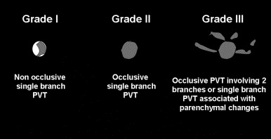
This is a retrospective cohort study of children admitted to the Toronto sick kids’ hospital NICU who had a diagnosis of portal vein thrombosis. They had 133 infants over 4 years, 73% of their infants had a UVC, and the majority of those who developed a thrombosis had a low or intrahepatic catheter position. Many of their patients progressed to lobar hepatic atrophy or portal hypertension (26% and 3% respectively). They also present a grading system, infants with more severe grades were more likely to develop those complications, and infants with mal-positioned UVCs were also more likely to develop complications. Many of their diagnosis were made when an abdominal ultrasound was performed for non-specific indications (babies with malformations or evaluation of hypertension) thrombocytopenia was relatively common at the time of the diagnosis. The authors could not find any indication that anticoagulation, which was given to about half of the babies, changed the outcome. They anticoagulated babies with a second thrombus, who had cardiac surgery, or who had a grade 3 thrombus.

This is their image showing the grading system.

portal-vein-thrombosis-grade

Shah PS, et al. A randomized, controlled trial of heparin versus placebo infusion to prolong the usability of peripherally placed percutaneous central venous catheters (PCVCs) in neonates: the HIP (Heparin Infusion for PCVC) study. Pediatrics. 2007;119(1):e284-91. This one is a little different, an RCT of heparin in picc lines, which showed that heparin prevents thrombosis of the catheter itself, but there is no evidence that it changes the rate of catheter associated central vein thrombosis (which occurred in 18% in each group, and were all non-occlusive). Which is very similar to the data from umbilical arterial catheters Barrington KJ. Umbilical artery catheters in the newborn: effects of heparin. Cochrane database of systematic reviews (Online). 2000(2):CD000507 heparin in UACs prolonged catheter patency, but did not affect arterial thrombosis rates. I also found one small trial of heparin for umbilical venous catheters (Unal S, et al. Heparin Infusion to Prevent Umbilical Venous Catheter Related Thrombosis in Neonates. Thrombosis Research. 2012;130(5):725-8.) there were only 19 babies in the heparin group (and 27 controls) with one control baby developing a thrombosis. Clearly not significant statistically, but woefully underpowered.

Park CK, et al. Neonatal central venous catheter thrombosis: diagnosis, management and outcome. Blood Coagul Fibrinolysis. 2014;25(2):97-106. This is a literature review, giving an overview of the topic and trying to synthesize the information from the mostly observational studies, many of which report different outcomes, and had different methodologies for detecting thrombi. They couldn’t find any reliable evidence that anticoagulation improves outcomes after a catheter associated thrombosis, they describe one study which reported the use of urokinase for catheter associated central venous thrombosis, about half of the clots resolved with this intervention but there were no controls.

Bohnhoff JC, et al. Treatment and follow-up of venous thrombosis in the neonatal intensive care unit: a retrospective study. J Perinatol. 2016. In this retrospective study the authors treated about 2/3 of the 26 central venous thromboses with low molecular weight heparin, the others had contra-indications to therapy. Doses needed were higher than the current recommendations especially for the preterm babies, some of the thromboses resolved, some of the babies were still treated after discharge. There were no hemorrhagic complications. There really isn’t any evidence from this study that the enoxaparin made a difference.

Saracco P, et al. Clinical Data of Neonatal Systemic Thrombosis. The Journal of pediatrics. 2016;171:60-6 e1. This is a report from an Italian registry, and includes 75 babies with systemic thrombosis, including arterial, venous and intracardiac thromboses. It really wasn’t very helpful for answering any of our clinical questions.

Finally, this article wasn’t discussed in our journal club, but it reports long-term outcomes from the sick kids cohort of portal vein thromboses (PVT). Morag I, et al. Childhood outcomes of neonates diagnosed with portal vein thrombosis. Journal of Paediatrics and Child Health. 2011;47(6):356-60. They found outcome data for 70 babies, and the duration of follow up was 2 to 8 years (LLA is hepatic left lobe atrophy). They conclude

…28% still had asymptomatic LLA in childhood, 7% had slowly progressive splenomegaly and 3% required shunting because of progression of portal hypertension. Ultrasonographic follow up was the most sensitive method in detecting progression associated with neonatal PVT. Until more data are available, periodic long-term ultrasonographic follow up should be considered for neonates diagnosed with PVT.

So what does this all mean? It is clear that central venous catheters can lead to central vein thrombosis, the major risk factors appear to be malposition of an umbilical venous catheter in the portal vein branch, and probably polycythaemia. Some children develop long-term complications of a thrombosis, with as many as 2 to 3% perhaps having portal hypertension, if the progressive splenomegaly is a sign of incipient portal hypertension then the incidence may be much higher. The anatomy of the region is complicated, and in fact looking for an image on google, representations of the anatomy are often wrong. This is a diagrammatic representation which I found here.

umbilical-vein-anatomy

Malpositioned umbilical venous catheters therefore often pass into the left branch of the portal vein, which may then become thrombosed decreasing perfusion of the left lobe of the liver. I am not sure why low catheters should have a higher risk of thrombosis than catheters in the ductus venosus or IVC, but that does seem to be the case in some studies, but not consistently. One idea for that association is that low catheters are often low because they won’t advance properly, and may have entered the portal vein during attempted insertion leading to endothelial damage and initiating thrombosis. It would be interesting to see if catheters intentionally inserted low (such as for resuscitation) also have an increased risk of thrombosis. Umbilical catheters left in place for more than 6 days also are more frequently associated with thrombosis, that agrees with the data from the RCT of UVC duration that I cited, where thrombosis recognition was after 5 days.

It isn’t clear to me that anticoagulation makes any difference, and I think that non-occlusive thrombi can probably be watched without therapy.In fact there is no good evidence that either heparin or LMWH improve outcomes for any site of thrombosis in the newborn as a very recent Cochrane review makes clear.

Occlusive and especially grade 3 thrombi have a higher risk of long-term adverse effects, but whether heparin, low molecular weight heparin (LMWH), or thrombolysis make any difference is not clear; LMWH can usually be given relatively safely, requires less monitoring as dose requirements are more predictable, and can be given intermittently. Thrombolysis always makes me anxious, I think it should be reserved for the most problematic thrombi, such as intracardiac cases.

So here are a few propositions, some are obvious some are speculative:

  1. Umbilical venous catheters should not be left in the portal vein.
  2. Umbilical venous catheters which are malpositioned (including low catheters) should be removed as soon as practicable
  3. Malpositioned UVCs should be followed by ultrasound evaluation of the portal venous system after removal
  4. Correctly positioned UVCs should be removed after about a week
  5. I question if all UVCs should be followed with ultrasound after removal or not, I think more prospective observational data are required. Perhaps well positioned catheters in non-polycythemic babies that are removed before 7 days might not need an ultrasound.
  6. Non-occlusive thrombi can be observed without anticoagulation
  7. Occlusive thrombi of grade 2 can probably be observed without therapy, but an RCT of LMWH would be very useful.
  8. Occlusive thrombi of grade 3, an RCT comparing no therapy, LMWH and thrombolysis (with rTPA) is needed, with long-term follow-up.
  9. Thrombi which are potentially dangerous in the short term (such as intracardiac) should be treated with rTPA
  10. For peripherally inserted central catheters no routine ultrasound is required (although frequent small thrombi are found, large clinically important thrombi  are rare)

I just searched clinicaltrials.gov and there doesn’t seem to be any relevant controlled trials of heparin or LMWH in the newborn. Off-label use of the LMWHs is quite frequent, so an RCT or 2 would be feasible within a research network.

Posted in Neonatal Research | Tagged , | 2 Comments

Inhaled steroids for BPD?

In a recent post I promoted a new Barrington rule for meta-analysis (it probably already existed somewhere but I am claiming priority anyway, it’s a “post-truth” kind of thing). The rule was that in a systematic review you shouldn’t pool data from 2 (or more) studies if it was impossible to be eligible for all the studies, in other words, if there is no overlap in the eligibility criteria. For example in the systematic review of inhaled NO in the preterm, there were studies of early rescue use for critically ill babies, and studies of much later use in relatively stable babies. Combining all those studies makes little sense as it doesn’t answer any clinically useful question. If you want to know about the proven effects of early use of inhaled NO for a 12 hour old baby who is needing 100% oxygen, then adding into the analysis studies of babies who were studied because they were on CPAP at 2 weeks of age is really unhelpful, and will only muddy the waters.

Which is why I really don’t understand the rationale for mixing together several of the studies in this new meta-analysis and systematic review

Shinwell ES, et al. Inhaled Corticosteroids for Bronchopulmonary Dysplasia: A Meta-analysis. Pediatrics. 2016.

The authors have included trials that studied :

(1) preterm infants of gestational age 22 0/7 to 36 6/7 weeks considered to be at risk for BPD, including both ventilated and nonventilated infants and (2) an RCT comparing any Inhaled Corticosteroid versus control (placebo or no treatment) at any dose and any duration of treatment and administered either by a metered-dose inhaler or by nebulization.


Which means they included studies with very early enrollment studying prophylaxis, other studies enrolling at 2 weeks of age of babies still intubated and with early BPD changes, and even one study that enrolled babies at 14 weeks of age who had already failed extubation and were needing more than 40% oxygen. I can’t imagine a clinical situation in which it would be useful to know the effects of inhaled steroids from all of those studies pooled data.

Most of the 16 included studies were tiny, with only 2 enrolling more than 100 babies, and most being around 30. Meta-analyzing multiple small studies risks inflating effect sizes. That isn’t the SR authors fault, but it is a problem with many neonatal systematic reviews.

The major primary outcome of the meta-analysis of Death or BPD at 36 weeks GA was based on 6 studies which are mostly somewhat similar to each other, and enrolled babies in the first 3 days of age (either on day one or on day 3) except for the study by Cole, which entered babies between 3 and 14 days of age. Bassler’s trial was a trial of prophylaxis in any baby needing assisted ventilation, whereas the others mostly needed some severity of illness criterion.

f1-large4

As I posted at the time that Bassler’s study was published that outcome is quite problematic, lung injury is a continuum, and forcing it into a dichotomous outcome for simplicity of reporting or analysis is rather unhelpful. Oxygen stopped at 35 weeks and 5 days, = no BPD, oxygen stopped 2 days later = BPD, even though the baby out of oxygen just before 36 weeks may have more signs of tachypnoea, retractions etc. Our recent publication (Janvier A, et al. Measuring and communicating meaningful outcomes in neonatology: A family perspective. Semin Perinatol. 2016) discusses this in some detail, and noted:

Many pulmonary outcomes having a negative impact on neonates and their families, such as rehospitalisation, needing oxygen therapy at home, and visits to the emergency department, are not systematically reported. A description of the impacts of the lung injury beyond a dichotomous BPD outcome would be much more useful, both to parents and to the health care professionals who care for them.

Although it is possible that very early inhaled steroids really do reduce oxygen requirements at 36 weeks, I am not sure this systematic review meta-analysis helps us know.

If you have a very preterm baby who is on respiratory support on the first day of life, and you want to know whether you should give inhaled steroids to reduce lung injury then the results of the studies in that Forest plot above from Yong, Fok and Bassler are relevant. In contrast, if the baby is not extubated by day 3 then the other studies are relevant, (Cole, Jangaard, and Merx). And even with all the data from both groups of studies that have non-overlapping eligibility criteria, the 95% confidence intervals are very close to 1.0.

So this isn’t really conclusive data, even about that outcome, death or BPD (of note there is no hint of an effect on death, the part of that outcome which is affected is the need for oxygen at 36 weeks), and as I said, I really don’t think it’s that important an outcome, unless, in this group, the diagnosis of BPD reflects worse pulmonary disease-related quality of life. What is now called moderate or worse BPD (i.e. oxygen for at least 28 days and still needing oxygen at 6 weeks) is associated with slightly worse long-term pulmonary function during the first year than mild BPD or no BPD, the differences are, however, not enormous. But just because a particular intervention decreases O2 requirements at 36 weeks doesn’t necessarily mean that those other clinically important outcomes (hospital re-admission, chronic pulmonary medication use etc) will be affected also. What I mean is, that steroids may reduce inflammation and have multiple effects on the lung, some of which might be negative, but unless they improve long-term lung function the baby hasn’t necessarily benefited.

I hope this Systematic review doesn’t lead to widespread use of a therapy for which the data are currently very limited, the excellent NEUROSIS trial will hopefully provide data about longer term pulmonary outcomes of their babies, to know if there is a benefit that will have an impact on the babies’ lives, and their families.


Posted in Neonatal Research | Tagged , , | Leave a comment

Toxicity of reflux medications in infancy

Before I leave the topic for at least a few days, I thought I would discuss data about the toxicity of reflux medications in older children. Most of my ‘toxic placebo’ comments have been about studies in preterm infants, and one of the major secondary effects of anti-acid medications in preterm infants is an increase in necrotizing enterocolitis and in systemic sepsis, neither of which are likely to be very common in older children. But there is in fact, also good evidence that anti-acid medications increase infections, at least respiratory infections, in older children.

Orenstein SR, et al. Multicenter, Double-Blind, Randomized, Placebo-Controlled Trial Assessing the Efficacy and Safety of Proton Pump Inhibitor Lansoprazole in Infants with Symptoms of Gastroesophageal Reflux Disease. The Journal of pediatrics. 2009;154(4):514-20.e4.  160 infants less than a year of age were randomized to lansoprazole or placebo if they had “symptoms of GERD”. There was no improvement in symptoms or any of the primary or secondary outcomes. There was however, an increase in serious adverse effects from 2% to 12%, the most common of which were lower respiratory infections.

Writing Committee for the American Lung Association Asthma Clinical Research Centers. Lansoprazole for children with poorly controlled asthma: a randomized controlled trial. JAMA. 2012;307(4):373-81. This multicenter trial was performed because of a widespread belief that acid reflux was responsible for triggering episodes of asthma, and anti-acid medications were frequently prescribed. Over 300 children were randomized (aged 6 to 17), and the proton pump inhibitor did absolutely nothing good. There was no improvement in any symptom, or any primary or secondary outcome. In contrast the children on the active drug had more respiratory infections (number needed to harm = 7) had more sore throats and had more than twice as many episodes of bronchitis (NNH=14).

The data from these 2 RCTs confirm  a prospective non-randomized study in 100 infants started on anti-acid drugs by pediatric gastroenterologists, both ranitidine and omeprazole were studied. There were significant increases in both gastroenteritis (from 20% to 47%) and pneumonia during the 4 months of treatment compared to the period before, and compared to the (non-randomized) controls. An observational study in adults also showed an increased risk of community acquired pneumonia.

So acid suppression doesn’t work for symptoms attributed to GERD  in older infants either, and the medications increase risks of infectious disease in infants, children and adults.

Metoclopramide has a poor safety profile with dystonic reactions, oculogyric crisis, irritability, drowsiness, emesis and apnoea occuring in 9 to 15% of patients, it also has not been shown to improve reflux, and indeed in a neonatal mammalian model, didn’t even have prokinetic effects.

Domperidone worsens GE reflux in newborns and has no beneficial effect in older children, it may prolong the QT interval; it is not thought to cross the blood brain barrier in older children, but the relatively ‘leaky’ BBB in babies might be another matter entirely.

So the data about efficacy and safety are very similar in older infants as in preterm babies: no good evidence of any benefit of any of the medications, and reliable evidence of harm.

They are still toxic placebos after hospital discharge.


Posted in Neonatal Research | Tagged , | 2 Comments

Anti-reflux medications in preterm infants; a whole cornucopia of toxic placebos.

I have written frequently about this on my blog in the past, and even written a book chapter about the subject (in the book titled “Nutrition for the Preterm Neonate” edited by Sanjay Patole). I recently mentioned the subject in my comments on the AAP choosing wisely recommendations.

The reason for posting about this now is a new publication, from a multi-site network of pediatric primary care practitioners that indicates that 37% of the babies born before 36 weeks received medication for reflux in their first year of life, most of which was started after NICU discharge. (D’Agostino JA, et al. Use of Gastroesophageal Reflux Medications in Premature Infants After NICU Discharge. Pediatrics. 2016). 40% of the treated babies received 2 medications, usually simultaneously, very often the 2 medications were both acid blockers, and some poor babies were getting 3 different acid blockers.

If you click on Gastro-oesophageal reflux in the word cloud at the left of this post you will be taken to a series of posts that address the issues.

But I will summarize:

Almost all babies have reflux, and almost all regurgitate occasionally, so we need to define what is GERD (GORD in England and anywhere else they know how to spell properly) or gastro-(o)esophageal reflux disease:
that is actually more difficult than you might expect, but basically that term means disease caused by gastro-oesophageal reflux, and includes changes caused by intestinal contents refluxing into the oesophagus or higher. Many articles about GER have diagnosed reflux by a percentage of time with acid in the oesophagus that is more than the 90th -percentile, which of course would mean that 10% of babies have ‘pathological’ reflux. But does that identify those babies that have reflux disease? Clearly not.

Can we clinically diagnose reflux? Basically no.

In the NICU there are no clinical signs which have been shown to differentiate between babies that have “excessive” reflux and those without, and one study with pH metry and video monitoring found just as many “reflux specific” clinical signs when the infant was not refluxing as when they were. Another study found that there was no correlation in the scores on 2 different supposedly validated scoring schemes for reflux, and another study showed that anti-acid medications had no impact on the symptoms that were attributed to reflux.

There are of course 2 broad categories of agents that are used for reflux, those which are given to reduce the frequency or duration of reflux events, and those which are given to inhibit acid production and hopefully reduce the adverse effects of reflux.

The first category consists of metoclopramide, cisapride, domperidone and erythromycin, (apparently now there are even people using bethanechol). All of these agents are toxic placebos, or toxic and worse than placebo, (that is both metoclopramide and domperidone have been shown to increase reflux) as you will see from the blog posts about each of them. Indeed as these are all prokinetic agents (although cisapride may not be prokinetic in the newborn) it is not too surprising that they may not improve, or may actually worsen reflux; increasing stomach emptying can easily send the contents in both directions!

Suppressing acid production is neither effective nor safe. It does not decrease symptoms that are attributed to reflux, and is associated with multiple complications. This is true whether you use good old ranitidine, or the latest expensive PPI.

I don’t understand why pediatricians would treat so many ex-preterm infants with agents that are ineffective and have secondary effects. Their use in the NICU has I think been decreasing, but much care of former preterm infants may well be delivered by general pediatricians who do not have the same approach as we do.

Time for more education, and perhaps more regulation, of drugs that have not been adequately tested in this population, are of uncertain efficacy, and have important secondary effects.

In the meantime, if you have a baby where you are convinced that their symptoms are caused by acid reflux, and you can’t stop yourself from prescribing an acid-blocking drug, the least worst harm you could do would be perhaps a “therapeutic trial”. Which means that after a few days if there is no improvement, stop the therapy. If there is an improvement, you should also stop the therapy, to see if the change is just due to the baby getting older, and if the symptoms recur, then you have a reasonable indication for continuing the treatment.

Posted in Neonatal Research | Tagged | 1 Comment

Neonatal Sepsis after Chorioamnionitis, what to do about healthy appearing newborns

In 2007, when I was chair of the CPS Fetus and Newborn Committee, we published a guideline regarding the approach to term and late preterm infants with perinatal risk factors for sepsis. Obviously any infant with clinical signs consistent with sepsis needs immediate work up and antibiotics, but the management of infants with risk factors for sepsis and no clinical signs evident was the focus of that guideline.

This is what we said about chorioamnionitis, based on what we thought was the most reliable literature:

The risk of sepsis (which may be due to a variety of different organisms, including GBS, E coli and other Gram-negative organisms) in an infant whose mother had definite chorioamnionitis is approximately 8%, and is approximately 3% to 4% if ‘possible’ and ‘definite’ chorioamnionitis are considered together [31][32] (evidence level 2b); among all mothers with fever, the incidence is 2% to 6% depending on the height of the fever [31] (evidence level 2b). Infants who do not have signs at birth are unlikely to develop sepsis, the odds ratio for sepsis among infants who are well at birth is 0.26 (95% CI 0.11 to 0.63) [31]. The incidence of invasive infection in the present study in an initially well-appearing infant with a maternal history of fever or chorioamnionitis was less than 2%, and this is confirmed by other data [33] (evidence level 2b). Therefore, it seems reasonable to perform a CBC and closely observe such an infant, and to only perform a full diagnostic evaluation and treat with antibiotics if the CBC is strongly suggestive of infection (low total WBC count) or if clinical signs develop. A requirement for extensive resuscitation at birth should be considered a sign of possible infection in such infants [32][33].

At that time the CDC guidelines recommended cultures and empiric antibiotics for all such infants, the CDC guidelines were updated in 2010, and continue to recommend the same thing, with the clarification that you should talk to the obstetricians (always a good idea!)

Well-appearing newborns whose mothers had suspected chorioamnionitis should undergo a limited evaluation and receive antibiotic therapy pending culture results (AII). The evaluation should include a blood culture and a CBC including white blood cell differential and platelet count; no chest radiograph or lumbar puncture is needed. Consultation with obstetric providers to assess whether chorioamnionitis was suspected is important to determine neonatal management (CIII).

Is there anything new recently? I think you all know the answer to that question.

Braun D, et al. Low Rate of Perinatal Sepsis in Term Infants of Mothers with Chorioamnionitis. Amer J Perinatol. 2016;33(02):143-50. This database analysis from Kaiser Permanent Southern California found an incidence of maternal fever in labour (38 degrees or more) at or after 35 weeks to be 9% and chorioamnionitis (based on ICD-9 codes from discharge data) to be 4%, chorioamnionitis based on a fever of 38 degrees followed by antibiotic treatment was 5%. There were around 30,000 deliveries in this cohort and in total there were 19 babies with culture positive early onset sepsis, 14 were symptomatic and 5 had bacteremia without clinical signs. That gives an overall incidence of culture positive symptomatic sepsis of 0.45 per 1000, and of culture positive bacteremia without clinical signs of 0.16 per 1000. Among mothers without fever the rate of sepsis was 0.5 per 1000, if they had fever without chorioamnionitis it was 0.6 per 1000, and if they had chorioamnionitis it was 4 per 1000.

One interesting thing in this study is that many physicians do not follow the CDC guidelines, the rate of neonatal treatment with antibiotics after maternal chorio, which should be close to 100%, ranged from 7 to 76% in different hospitals.

The authors don’t say how many of the babies without clinical signs were from the maternal chorio group, there were 5 babies who did not have clinical signs, if all of them were from mothers with chorioamnionitis (which I think is unlikely) then you would have to treat 250 clinically well infants after maternal chorioamnionitis with antibiotics to be sure to cover 1 baby with bacteremia. Of course if some of those babies were symptomatic, then you would have to treat many more without clinical signs for each baby infected, for example if the proportion of asymptomatic babies is the same as in the next study (about 1/3) then you would have to treat over 700 asymptomatic babies to cover that 1 with bacteremia.

The authors also calculated that if the CDC guidelines had been followed (for chorioamnionitis, but also for other indications for neonatal antibiotic treatment) then 8% of all the term and late preterm babies would have received 48 hours of antibiotics.

Another very large study Wortham JM, et al. Chorioamnionitis and Culture-Confirmed, Early-Onset Neonatal Infections. Pediatrics. 2016;137(1). instituted prospective surveillance for early onset sepsis among nearly 400,000 deliveries. In contrast to the other study they did not collect data from mothers whose babies did not get septic, so we don’t know overall incidence of chorioamnionitis in this study, and you can’t make some of the same calculations.
They found 389 cases of early onset, culture positive, infections, in the cohort which included both preterm and term babies. Eighty-one of those infections were in term babies (37 weeks and more; from 350,000 term deliveries) 58 of which had clinical signs at birth. Which leaves 23 term infants who had no signs at birth but had culture positive sepsis, from mothers with chorioamnionitis, and 6 preterm babies with the same combination. The authors include some babies, 1 at  term and 4 preterm, who only had histological evidence of chorioamnionitis, which doesn’t help our decision-making as you don’t know about those unless you did placental pathology and got the results back.

If the prevalence of chorioamnionitis is 4 to 5% (as in the first study), then there would have been about 16,000 cases of chorio among the term deliveries, which gives the incidence of early neonatal sepsis (81/16,000) of 5 per 1000 which is very similar to the Kaiser Permanent data. We know 23 were without signs at birth (but one of those would not have had a diagnosis of chorio) so to ensure that 22 those asymptomatic babies with sepsis received early treatment, you would have to screen, culture and treat 16,000 babies, which is a number needed to treat of over 700.

I think that is too many. NICE in the UK seems to have a similar opinion in their guideline from 2012, their guidance is a little more complicated, but goes like this (the links should work to take you to the tables):

Use the following framework based on risk factors and clinical indicators, including red flags (see tables 1 and 2), to direct antibiotic management decisions:

  • In babies with any red flags, or with two or more ‘non-red flag’ risk factors or clinical indicators (see tables 1 and 2), perform investigations (see recommendations 1.5.1.1–1.5.1.3) and start antibiotic treatment. Do not delay starting antibiotics pending the test results (see recommendations 1.6.1.1–1.6.1.3).
  • In babies without red flags and only one risk factor or one clinical indicator, using clinical judgement, consider:
    • whether it is safe to withhold antibiotics, and
    • whether it is necessary to monitor the baby’s vital signs and clinical condition – if monitoring is required continue it for at least 12 hours (at 0, 1 and 2 hours and then 2-hourly for 10 hours).


In their scheme chorioamnionitis is a risk factor, but not a “red flag”, so if the baby does not have any of the clinical indicators they would be observed, but not treated.

As I said I think that an NNT of over 700 is too many, but others may not agree, specifically the parents may not agree. Shouldn’t they be involved in that decision? A decision aid may help them to decide, with the medical team, between one or multiple IV installations, antibiotics for 36 hours (you can stop them before the 48 hour dose if cultures are negative) and hospitalisation for at least that long. Depending on how your hospital is organized term newborns on antibiotics may also not be in the room with the mother. The contrasting choice is close observation with the option of starting antibiotics later if clinical signs appear (which happened in 18 of the 23 initially asymptomatic term babies with positive cultures). Also there is no difference in mortality (there was one death in the Wortham study, a baby who died soon after cultures and never had antibiotics, 2 deaths in the Braun study both of whom were symptomatic) shown in these recent studies.

Very frequent use of antibiotics of course affects colonization and resistance patterns in an environment, and will affect the development of the infant’s microbiome, perhaps for many months or years.

I think in these days of shared decision-making and family centered care, when a term baby is born after maternal chorioamnionitis but is clinically well, we should inform the parents that the baby has a very small risk of having an infection, (1 in several hundreds) and that there are 2 options. We should also organize our care so that both close repeated observation, and/or antibiotic administration, can be performed in a mother and infant room, without interfering with breastfeeding and the evolution of the new family, and then give the parents a big place in that decision.


Posted in Clinical Practice Guidelines, Neonatal Research | Tagged , , | 1 Comment

Podcast starring Annie Janvier: the harms of a gestational age label

The Archives of Disease in Childhood (and several other BMJ journals) have a series of podcasts, most of them seem to be interviews between a member of the editorial board and an author of a recent article. In this new one Jonathan Davis, one of the associate editors of the fetal and neonatal edition, interviews Annie Janvier about an editorial that she has just written for the journal. (Delivery room practices for extremely preterm infants: the harms of the gestational age label)

I already posted about the associated article and mentioned the editorial, but the podcast is 24 minutes long and goes into more detail about some of the issues.

Posted in Neonatal Research | Leave a comment