PAS 2023, some supplemental selected stuff

As usual, the annual meeting of PAS had too many things going on simultaneously to be able to get to all the interesting looking neonatal research. But here are a few things, that were of interest to me, and which I either got to in person or wanted to.

How much CPAP after extubation?

The Éclat trial (happy to see a French trial acronym) studied babies <28 weeks who had received surfactant and were on caffeine and were about to be extubated. They were randomized to either standard CPAP pressures after their first extubation (6-8 cmH2O) or higher pressures (9-11). The primary outcome was extubation failure within 7 days, which was defined as the FiO2 increasing by 20%, or a resp acidosis (PCO2 >60 and pH <7.20), bad apneas or urgent need for re-intubation. This is their poster (from the PAS website), which shows there were many fewer extubation failures using higher CPAP, and no difference in other adverse outcomes.

With 69 per group they were not powered for many other outcomes, and it is interesting that the incidence of BPD is very high in both groups, but I don’t know what definition this was using, I would guess it included mild BPD.

Would you like a side order of budesonide with that surfactant?

The results of the Taiwanese trial (sadly lacking an acronym) which randomized infants to surfactant alone (Curosurf) or poractant with budesonide (0.25 mg/kg/dose) and allowed up to 3 surfactant doses in each group, were presented. To be eligible babies were <1500 g at birth and required intubation in the delivery room or within 4 hours after birth. There were slightly over 300 babies in the trial, and the primary outcome was “BPD or death”. The average GA was about 27.5 weeks in each group. Mortality was similar in the 2 groups at close to 10%. 60% of the surviving controls developed BPD, and 38% of the surviving budesonide babies, with, also, more mild cases in the steroid group, and more severe cases in the controls.

It looks like the steroids were more effective in the larger more mature babies, secondary analysis by weight stratified subgroup doesn’t shown an effect in the <750 g babies, but there were less than 40 per group who were this small, and very little power. Blood pressures were higher in the treated group, suggesting systemic absorption, which you would expect. Alan Jobe has shown in preterm lambs that the majority of administered budesonide enters the systemic circulation, less than half being left in the lungs. I didn’t see any data on late-onset sepsis, and long term follow up will be important, as it is very early steroid use which has been most strongly associated with cerebral palsy in previous studies of systemic use.

None of the previous trials of steroids which have shown a decrease in BPD, and which have followed the babies after discharge, have shown improved long term respiratory health. Long term respiratory outcomes will be essential.

This looks encouraging, and I know some centres are doing this already, we will discuss in my centre, but I’m not sure I’m quite ready to expose every intubated VLBW to steroids on day one. Awaiting the PLUSS trial with bated breath.

Optimistic for long term pulmonary outcomes

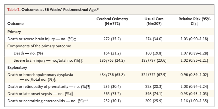
Peter Dargaville presented the long term pulmonary and developmental outcomes of the OPTIMIST trial of minimally invasive surfactant treatment. In that trial 488 babies of 25 to 28 weeks gestation were randomized to MIST or ongoing CPAP or nasal IMV (PEEP of 5 to 8) if they needed 30% oxygen or more in the first 6 h of life. The primary outcome was “death or BPD”; there were slightly more deaths in the MIST group (10% vs 8%) but less BPD among survivors, 37% vs 45%. “Death or BPD” was therefore a bit less frequent with MIST (44% vs 50%) but was “not statistically significant”.

The follow up of that trial up to 2 years of age gave this CONSORT flow diagram, showing that up to 2 years the mortality was very similar. For various reasons, especially COVID, in person follow up was only the minority, but they had on-line questionnaires filled in by parents, and all the developmental outcomes were similar between groups. The proportion of babies with language or cognitive delay was identical between groups.

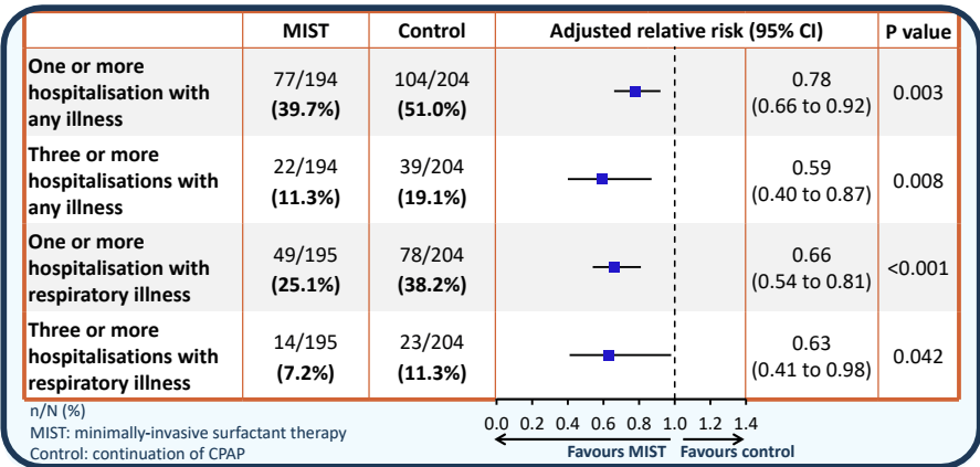
In contrast the respiratory outcomes all look better in the MIST group.

There were questions about whether this was really a trial of early versus late surfactant, but as Peter rightly said, it was not! Control group babies did not all get surfactant, babies in both groups were intubated for surfactant if they reached 45% (MIST was not allowed) and after intubation further therapy was according to usual practice, so INSURE, or more slow weaning, or whatever your usual was were OK. Most babies were expected to be on caffeine, but it was not mandated by the protocol.

This is one of very few studies of respiratory interventions which seems to show clear long term respiratory benefits, beyond just the presence of a BPD diagnosis. 72% of the controls got intubated within 72 hours of birth, compared to 37% of the MIST babies.

MIST in the delivery room, with coffee!

Anup Katheria presented another trial of thin catheter administration of surfactant, this time being randomized in the first hour of life, once babies of 24 to 29 weeks were stabilised on CPAP, and had received caffeine. Controls also received caffeine, but no surfactant, 180 babies were randomized and the main outcome was need for intubation, determined by needing over 40% O2, or a respiratory acidosis or bad apneas. Babies with less-invasive surfactant and caffeine required intubation in the first 72 hours of life 23% of the time, compared to 53% of the time for the caffeine only group. The surfactant group did get their caffeine a bit earlier than the controls (at 52 minutes of age compared to 70 minutes). Death before discharge was rare in this study (only 3 babies died, all in the control group), and there seems to be less BPD in the surfactant group, 26% vs 39%, although the study was not designed to have power for the BPD diagnosis, it looks like there probably was an effect.

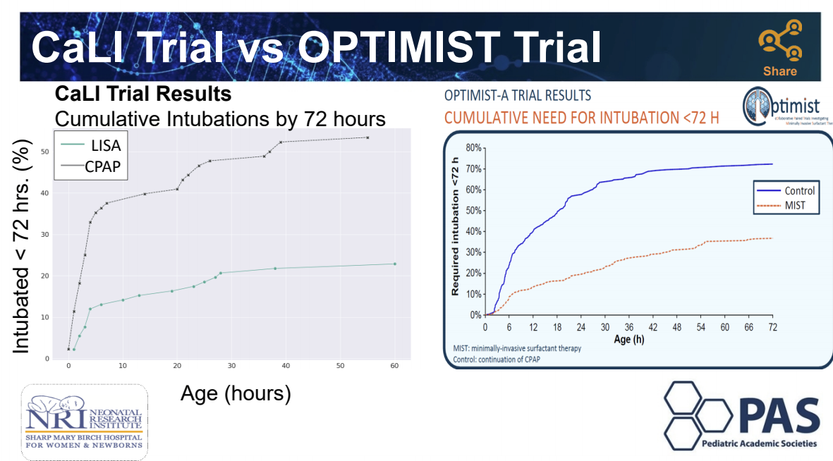
Anup Katheria helpfully listed the differences between his study and OPTIMIST

I really hate the idea of performing laryngoscopy without analgesia, and sedative premedications were not allowed in the OPTIMIST trial. I don’t know if they were allowed in CaLI, but if we can avoid intubation completely in a large number of LISA/MIST babies, which these survival curves below demonstrate dramatically, and therefore avoid prolonged desaturations, bradycardias and multiple intubation attempts, then maybe an un-premedicated less-invasive surfactant in early life will reduce, overall, the amount of pain experienced.

Putting things together

It is starting to look like very early minimally-invasive surfactant administration, perhaps routinely in the DR, and with very early caffeine administration, is the way to go. Not because it decreases “BPD” but because it seems that long term respiratory health is improved by avoiding intubation in the first few days of life (long term outcomes of SUPPORT and now OPTIMIST)

Perhaps early less-invasive surfactant should be co-administered with budesonide, but we should carefully consider that the NEUROSIS trial, of repeated budesonide inhalations starting during the first 24 hours, showed a small excess of mortality in the budesonide group. OPTIMIST also shows a small excess of mortality with budesonide. In addition, budesonide when mixed with surfactant enters the systemic circulation, and very early systemic steroids in preterm infants increase motor delay and cerebral palsy.

Of all the previous steroid trials that have shown reduced BPD and have also reported long term respiratory outcomes, I don’t think there is a single one which has shown improved longer term respiratory health.

Of course those studies are all contaminated by frequent treatment of control babies with steroids, so long term outcomes may be no different for that reason. On the other hand, potent systemic Steroids may decrease inflammation and thus lead to fewer babies needing oxygen at 36 weeks, but they also impair lung growth and interfere with lung development. I would like to see some evidence that long term respiratory health is improved before treating all babies receiving surfactant with budesonide, not to mention some evidence that there are no adverse effects on neurologic or developmental outcomes.

Posted in Neonatal Research | Tagged , , , , | 2 Comments

PAS 2023, some selected stuff

Hernia repair timing

One trial which will should have an impact on the care of newborn infants is the trial of early versus late hernia repair. 40 neonatal centres in the USA randomized 320 preterm infants to inguinal hernia repair before discharge (between 37 and 42 weeks, if I remember correctly) or later repair after 55 weeks PMA (55-60 weeks according to the record on clinicaltrials.gov). That record also notes a sample size of 600, and notes that a Bayesian analysis of the data will be the primary method. The primary outcome was a composite of adverse events, which I can’t now find details for but included things like post-operative apnea, requiring intubation post-op, and incarceration of the hernia.

The justification for pre-discharge repair has always been that incarceration was relatively common, and we should repair the hernia before discharge so that doesn’t happen. Incarceration did occur in 4.5% of the late repair group, it was defined as needing surgery, sedatives or a surgeon to reduce the hernia. There were a few incarcerations in the early treatment group as well.

Overall there were more adverse events in the early surgery group, 26%, than the late group, 18%, which made it highly likely (96% probability) that late surgery led to fewer adverse events. The total number of hospital days after randomization was also shorter in the late repair group. One somewhat unexpected finding was that 10% of the hernias resolved in the late group. Apparently this has been suggested before (many years ago), and was probably a real finding, as the presence of most of the hernias was confirmed independently.

Some of our families live hundreds or thousands of kilometres away, and re-admission for later hernia repair may be more of a problem for them. But for the majority of families who live with easy reach of my hospital, it looks like we should probably wait for 3 months post-discharge before fixing their hernias.

The only real disappointment of this trial is the lack of a catchy acronym, it could have been HIDE (Hernias of the Inguinal region, Delayed vs Early repair) or some combination of S (for surgery) HI and T (for trial)…

Premies are cool enough

A randomized trial of cooling for asphyxiated preterm infants who have not been well studied in previous trials (33 to 35 weeks gestation) but who sometimes get cooled, because of “mission creep”, was performed by the NICHD network.

The calculation of sample size was interesting, as they designed the trial to get the largest sample which they considered feasible within a reasonable time, and planned a Bayesian analysis of the trial. They planned to enrol 168 babies, and achieved that, and care was identical to that for more mature babies, with careful normothermia in the controls, and 33.5 degree target temperature in the hypothermia group. Cooled babies were more likely to become hypoglycaemic, and more likely to have major bleeding, but most importantly more likely to die, 21% vs 15% with a Bayesian calculation of a 77% probability of a real increase in mortality with cooling compared to normothermia.

The primary outcome was death or “disability”, which was a Bayley 3 cognitive score <85 or GMFCS >2 or a seizure disorder or deafness. The primary outcome was very similar between groups, with a neutral probability on Bayesian analysis of being better or worse.

This is probably the best evidence we will get for the efficacity of therapeutic hypothermia in late preterm infants, and strongly suggests that it is not effective, and may be harmful.

Mild HIE, to cool or not to cool, that is the question.

From Imperial College London a combined report of 2 trials targeting term babies with signs of mild encephalopathy. They had to have a clinical examination, in the mild category, and a normal aEEG. The trials includedonewith babies randomized before 6 hours, who were then treated with normothermia or standard hypothermia, and a second group of babies who were already cooled, and who were >6 hours of age, they were randomized to shorter or standard hypothermia.

The trials are quite small, and so far only short term outcomes and MRI were available. Cooled babies were more likely to be intubated and to need inotropes. They also had longer hospital stays. As for the MRI results, they appear to be worse in the cooled groups than the normothermic infants.

As I wrote previously MRIs are very poorly predictive of outcomes on an individual basis, but there are overall correlations in groups of babies between poorer outcomes and worse MRI scores.

Therapeutic hypothermia for mild HIE has been growing in frequency, and I have been guilty of starting it when I wasn’t sure whether to, or not. I felt from previous data that it was probably safe, and possibly effective. I may have to revise that opinion, but clinical examination may change prior to the time limit for cooling, and babies with normal or mild encephalopathy may deteriorate. I think based on this data that we should be cautious cooling babies who are clinically in the mild category, ensure that they are re-examined at 5.5 hours and have aEEG or EEG. If the examination only shows signs of mild encephalopathy, and the electricity is normal, then scrupulous maintenance of normothermia and supportive care is indicated.

Posted in Neonatal Research | Tagged , , , | 2 Comments

I might have already mentioned this; CRPs are CRaP!

Acute phase reactants increase for many different reasons, such as being born. Other causes of increased CRP include maternal antibiotic prophylaxis, TTN, HMD, Surfactant treatment, meconium aspiration syndrome, prolonged rupture of membranes, HIE, higher birthweight, gastroschisis, and probably someone scowling at the baby,

They do not rise instantly when a baby is infected, but take a variable amount of time to go up. Specificity and sensitivity for bacterial sepsis are both very poor, we should stop using them for late onset sepsis. A Cochrane review, latest update 2019, revealed a sensitivity of 0.74 and a specificity of 0.62 for late onset sepsis, which, combined with the fact that the majority of screens for late onset sepsis are negative, means that the PPV for a raised CRP is less than 20%, and the NPV of a low CRP is about 5%

For early onset sepsis, there are still many places where they are included in initial evaluation, and repeated CRPs may be used to decide on continuing antibiotic treatment. In some studies an increased CRP is used to define late-onset sepsis, even in well-appearing babies. Which leads to some interesting circular arguments about who should be treated with prolonged antibiotic courses.

This latest study (Dhudasia MB, et al. Diagnostic performance and patient outcomes with C-reactive protein use in early-onset sepsis evaluations. J Pediatr. 2023) is about babies admitted to 2 NICUs. It covers periods with routine CRP measurement as part of an EOS evaluation, and then a second period when CRPs were no longer routine, they were not banned, but became progressively less frequently requested.

They give results from 2009-2014, where CRP was routine “10,134 infants were admitted; 9,103 (89.8%) had CRP and 7,549 (74.5%) had blood culture obtained within 3 days of birth. CRP obtained ±4 hours from blood culture had a sensitivity of 41.7%, specificity 89.9% and positive likelihood ratio 4.12 in diagnosis of EOS. When obtained 24-72 hours after blood culture, sensitivity of CRP increased (89.5%), but specificity (55.7%) and positive likelihood ratio (2.02) decreased”.

They then compare approximately equal periods of about 2 years each with (n=4,977) and without (n=5,135) routine use of CRP. Of note the later period was also the time when the centres started using the Kaiser EOS calculator for the term and late preterm babies, and stopped doing some sepsis evaluations in preterm babies with ultra-low risk delivery characteristics. “We observed lower rates of EOS evaluation (74.5% vs. 50.5%), antibiotic initiation (65.0% vs. 50.8%), and antibiotic prolongation in the absence of EOS (17.3% vs. 7.2%) in the later period”.

They also showed no difference in the incidence of EOS (about 2 per thousand), no difference in how long it took for septic babies to receive antibiotics, and no differences in clinical outcomes.

CRP results do not sufficiently discriminate between infected and non-infected babies to be useful. This is true for EOS and for LOS. Abandoning CRP use has no adverse impact, but reduces antibiotic exposure of non-infected babies.

Posted in Neonatal Research | Tagged , | 4 Comments

An ESCape route for infants suffering opiate withdrawal

The Finnegan score was developed and evaluated in the 1970’s as the first systematic way of monitoring babies going through perinatal drug withdrawal. Although it was an advance at the time, it was not developed using modern standards, and was never, I don’t believe, studied in a randomized trial, indeed it would be hard to know what you could have compared it to, other approaches were rather haphazard.

The Finnegan scoring system, and the treatment approaches based on it, became the default approach, and have been the standard of care for many years. The problem of neonatal drug withdrawal seems to be getting much more frequent, especially in the USA, reliable prevalence data are of course difficult to obtain, but 7% of US mothers report taking opioids during pregnancy, 1/5 of them report abusing opioids, rather than therapeutic use. The incidence of neonatal abstinence syndrome, NAS, nearly doubled between 2000 and 2017, and I think all indications are that it is still increasing, with 7 cases per 1000 births in 2017. I.E. nearly 1% of all births in the US.

In Canada in 2020 the incidence was about 6 per 1000 live births (not including Quebec who collect data separately). The opioid crisis in Canada is, generally speaking, worse in the west of the country and is spreading eastward, with the highest rates of overdose deaths in British Columbia, followed by Alberta then the prairies and Ontario, with far fewer cases as yet in Quebec.

With this enormous and increasing incidence, evidence-based methods for evaluating and intervening for these infants were required, and a new program, first reported in 2017 was developed, based on a simplified evaluation (which is where ESC comes from: is the infant able to eat, to sleep, and to be consoled), use of non-pharmacologic measures to calm the infant, reduced stimulation, skin to skin care, encouraging breast feeding, and limiting morphine use.

Several publications strongly suggested that the ESC approach was followed by a reduction in duration of hospital stays, and less opiate administration. This new publication (Young LW, et al. Eat, Sleep, Console Approach or Usual Care for Neonatal Opioid Withdrawal. N Engl J Med. 2023) reports the first large RCT, performed as a cluster randomized trial, with all the 26 centres switching from standard care to ESC, but doing so at times which were determined randomly; a “stepped wedge” design.

It would be easy to criticise this design, it is almost implicit in the design that ESC is preferable! No cluster was randomized to switch from ESC to standard care, which you could do if there was really equipoise. I think it shows that most people were already convinced that ESC was the better approach, but they wanted some scientifically valid way of confirming that. The design does give real-world information about the size of the impact of ESC, and allowed some post-discharge evaluation of safety.

The primary outcome of the trial was the age of being medically ready for discharge, defined as : “an age of at least 96 hours, a period of at least 48 hours without receipt of an opioid, at least 24 hours with no respiratory support and with 100% oral feeding, and at least 24 hours from initiation of maximum caloric density.” over 1300 babies were enrolled, and 837 were discharged when medically ready, and therefore contributed to the primary outcome.

There were therefore 468 who were discharged before they met this definition, almost all were either <96 hours of age (n=211), or it was <48 hours since their last opioid dose (n=231). The investigators therefore also evaluated a “modified definition of medical readiness for discharge” which included an age of at least 72 hours and at least 24 hours without receipt of an opioid. This definition could be evaluated for 1164 of the infants.

Just getting the babies home earlier, while a benefit of itself, is not necessarily an advantage if the babies need to be re-admitted for further NAS therapy, or for other related reasons, or if parental coping is affected and more babies have adverse outcomes. The babies were therefore followed for 3 months post-discharge for the following outcomes “(any acute or urgent care visit, emergency department visit, or hospital readmission), and a composite critical safety outcome at discharge and through 3 months of age (nonaccidental trauma or death).”

Follow-up of these babies may be very difficult, especially in the US health environment. “Outcomes after hospital discharge were assessed prospectively at 3 months of age by means of a review of electronic medical records (including linked medical records) and media review through a search of public records (e.g., news reports, obituaries, and registries)”.

The main results are below: as I mentioned, the primary outcome was assessed for 837 babies, but the actual mean length of hospital stay includes all the babies, as does the dramatic difference in the proportion who received morphine.

The modified definition occurred at 14.5 days in the usual care group, and 8,1 days in the ESC group.

For the 3 month outcomes :

About 2/3 of the hospitalisations in each group were possibly related to NAS.

This study should be the death-knell (whatever a “knell” is) for the Finnegan score. It is not particularly objective, nor clearly very useful for determining therapy. The simplified ESC system should rapidly become the standard of care, and the standard against which any innovations in NAS care should be tested.

As one example, there are several trials of buprenorphine for NAS, as an alternative to either morphine or methadone, those trials (such as this one) have generally used the Finnegan system, and have often shown shorter treatment with buprenoprhine than the alternative. The effect size of ESC is larger than that shown in those trials, in general. Maybe some of them should be redone, with ESC as the approach to care in both groups.

Posted in Neonatal Research | Tagged , , | Leave a comment

PAS meeting starting tomorrow. Panel Saturday afternoon!

For those of my readers going to PAS in Washington DC, I have put together a panel to discuss painful procedures in neonatal research projects, which starts at 2pm on Saturday, April 29, 2023. The stellar cast of speakers include the moderator John Lantos, who will introduce the topic and talk about research ethics and how painful interventions in children and babies fit into the ethical framework. I will give some examples of painful procedures in neonatal research projects, how they were justified, if at all, and try to create some guidelines for what might be acceptable, Ruth Grunau will talk about how pain adversely affects the developing brain, and Sunny Juul will talk about designing research projects that minimize additional pain.

I think it will be an important and informative session, hopefully with enough chairs for everyone who wants to attend. It is in the convention center 145 AB.

Posted in Neonatal Research | Leave a comment

What now for cerebral NIRS in the preterm?

Previous randomized trials of routine cerebral NIRS in preterm infants have shown that cerebral hypoxia can be reduced by responding to the signal, but in an underpowered follow up of a subgroup, there was no impact on long term outcomes. The same investigators, in an expanded collaborative of 70 NICUs studied the impact of routine monitoring of cerebral oxygenation on brain injury as shown on ultrasound up to 36 weeks PMA. Hansen ML, Pellicer A, Hyttel-Sorensen S, Ergenekon E, Szczapa T, Hagmann C, et al. Cerebral Oximetry Monitoring in Extremely Preterm Infants. New England Journal of Medicine. 2023;388(16):1501-11.

The definition of the outcome was “one or more of: intraventricular hemorrhage of grade 3 or 4, cystic periventricular leukomalacia, post-hemorrhagic ventricular dilatation, cerebellar hemorrhage, and cerebral atrophy” which are all defined more precisely in the supplemental material. One quibble I have with the definitions is of grade 4 IVH, defined as “parenchymal haemorrhagic infarction visible in the periventricular white matter”, what we actually see is echodensity, and it is not possible to really ascribe a mechanism of injury to an ultrasound appearance. As an upcoming review article that I co-authored will point out, periventricular echodenisities are not all strongly associated with poor long term outcomes. The extent, location and whether uni- or bi-lateral are all important.

This study was large enough (n=1600) to show moderately large differences in the incidence of ultrasound abnormalities, 90% power to show a reduction from 34 to 26% among infants <28 weeks GA. Monitoring had to start within 6 hours of birth and continue for 72 hours, a treatment algorithm was followed to protocolise responses to low cerebral saturations.

The results in the 2 groups were, basically, identical.

My big question is whether this is a surprise, I think that low cerebral oxygen saturations are due to a large number of different processes, and that there is not a clear link between low cerebral oxygen saturation and severe IVH or PVL. Only 29% of the babies in the oximetry group had a change in management because of low saturations, so you would only expect the intervention to have any impact on that 1/3 of the babies. PVL may occur after an episode of shock, but the strongest correlation is with perinatal inflammatory disorders, like chorioamnionitis.

I also think it is a real possibility that less cerebral hypoxia could improve long term outcomes without necessarily changing brain ultrasound findings, and I really hope that after this great effort to perform a really important trial, there will be long term follow up of the survivors.

For now there is no clear evidence that routine cerebral NIRS, and responding to low cerebral oxygenation, improves short or long term clinical outcomes in the preterm.

Posted in Neonatal Research | Tagged , , , , | Leave a comment

Survival after End-of-Life Discussions

Another of the articles that have been keeping me busy over the last couple of months has just been published. Boutillier B, Biran V, Janvier A, Barrington KJ. Survival and Long-term Outcomes of Children Who Survived After End-of-Life Decisions in a Neonatal Intensive Care Unit. Journal of Pediatrics. 2023:113422.

Discussions with parents about the progress of their baby, when we have concerns about their survival, or poor prognosis, are not infrequent in the NICU. We may decide to continue as before, or to limit interventions, or to withdraw some, or all, life sustaining interventions. One of our fellows, Beatrice Boutillier had collated the results of a large number of such discussions in a French tertiary NICU, and investigated the outcomes, who died, who survived after treatment limitation or withdrawal decisions, and the long term outcomes of those who survived. Annie Janvier and I collaborated with Beatrice, and with Valérie Biran, the chief of the NICU at Robert Debré hospital, to publish the data from their NICU.

There is very little other such information in the literature. Although there are several publications about the proportions of deaths that follow end of life discussions, many of those have collected data only on babies who died, there is little about those who survive after a decision to limit or withdraw care.

Although each case is unique, we squeezed them into general categories, using a revision of the categories of Eduard Verhagen et al. In some, all ICU care was withdrawn, including assisted ventilation, this could either be because it was thought there was little chance of survival in an unstable infant (category C), or because the long term prognosis for an acceptable quality of life was thought unlikely (category D). There were some infants where there was a decision to not escalate care, such as deciding to not go to the OR, or to not start an inotrope, or sometimes just to withhold CPR, these were category B. Those with withholding some interventions or limiting care were the babies most likely to survive, 18/29. But even among those with withdrawal of life-sustaining interventions, 4/41 unstable, and 12/94 stable infants survived.

All of the babies who survived to discharge required some medical follow up, and for many there were multiple specialties involved. Of the 34 who survived to 2 years, we had functional outcome information on 32, 8 of whom had outcomes between functionally normal, up to functional limitations with likely supervised living in the future, the remaining 24 were predicted to need help with activities of daily living or to be partially or totally dependent. We used the Glasgow Outcomes Score-Extended, as adapted for children (which I think gives a much better picture of the functional abilities of the children than a label of “NDI”).

The results point out the uncertainties inherent in our practice; even unstable babies that you think will likely die, who are so sick that you decide to withdraw their life-sustaining interventions, may still survive. After doing neonatology for over 40 years now (I know, I don’t look that old) I find myself often humbled by seeing things I would have thought impossible. Recently I looked after a baby with a pH <7.00 for 12 hours, (the baby was on extreme support, the parents weren’t ready to withdraw LST, so we continued while assuring good analgesia), who survived and is doing OK after discharge.

I am sometimes tempted to give up trying to predict anything! But I think it is better just to be transparent and honest and be open about the uncertainties of life, and of neonatology. We must always recognize that we are lousy at predicting survival, and even worse at predicting long term functional disability. Even with all the tools at our disposal, it is rare that we can say anything definitive.

One thing that doesn’t seem to help parents is to try and develop predictive models which give a percentage likelihood of survival or of poor long term outcomes. Although it is a different time of life, a recent study is relevant McDonnell SM, Basir MA, Yan K, Liegl MN, Windschitl PD. Effect of Presenting Survival Information as Text or Pictograph During Periviable Birth Counseling: A Randomized, Controlled Trial. Journal of Pediatrics. 2023. This study, using a vignette of a threatened delivery at 22 weeks gestation, showed that it didn’t make any difference how outcome data were presented (text or pictogram) to treatment decisions, it also showed that it didn’t make any difference what data were presented! Whether a 30% or 60% survival probability were revealed to a randomly selected group of 1000 women of child-bearing age didn’t make any difference to decisions, it didn’t even make any difference to what the participants thought the chance of survival was; when told the baby had a 30% chance of survival the participants thought the baby had a 68% chance of survival. Participants, however, thought that even with palliative care the baby was likely to survive (median 58% chance of survival), which does show that the presentation of the information, no matter how it was done, did not lead to the respondents having accurate knowledge of the results of palliative care.

The article is reviewed by a really nice editorial from Naomi Laventhal (Laventhal N. Negative Studies and the Future of Prenatal Counseling at the Margin of Gestational Viability. Journal of Pediatrics. 2023:113440.) who points out that the data are consistent with other studies, and that although there are several tools for calculating percentage survival and disability, there is no good data to show that such percentages are what parents want, or that it makes a difference to decisions made, even if they accept the figures they are given.

“These seemingly nihilistic study results are illuminating. Should we be spending our time trying to get better at sharing outcome probabilities with expectant parents if the probabilities aren’t what matter to them?”

I never give percentage survival figures, unless parents ask for them (which they almost never do), and, to be honest, I am not at all sure that the difference between 30% survival and 60% should make a difference in decision-making. You might as well say 50:50, and leave it at that for both cases. There is a reasonable chance of survival, and ICU care is probably worth a try, I would say, and most of the respondents in the article by McDonnell seem to say the same thing. It might be different if the chances were 1% vs 99%, but it is hard to think of a scenario which could be included in such a study where the chances would be 1% survival.

To return to the case series that we just published, some of the considerations in counselling in the NICU are similar. We can try and calculate survival rates, but each case is different, each individual baby can only be 100% survival or 0% survival, and parental (and physician) attitudes and values are probably more important to the outcomes of such decisions than percentages. We have to always remember that neonatologists are remarkably bad at predicting the future.

We should also remember that parents of surviving preterm infants almost never regret their life and death decisions. In another study that we published recently, Thivierge E, et al. Guilt and Regret Experienced by Parents of Children Born Extremely Preterm. Journal of Pediatrics. 2022, 113 of 248 parents of former preterm infants, seen in the follow up clinic, expressed some regrets about the NICU stay. None of them regretted decisions made to start or continue NICU care. We know from other data that decisional regret is more common after decisions to opt for palliative or comfort care alone. The regrets that parents did express were more associated with lack of self-care during the NICU stay, regrets about having a preterm baby (for which mothers often blamed themselves) and regrets related to their role as parents in the NICU. Addressing those issues could help to improve parental mental health after discharge.

Posted in Neonatal Research | Tagged | Leave a comment

Imaging after therapeutic hypothermia: when to do the MRI (if at all)

I had thought it was well settled that imaging soon after rewarming was as good for predicting outcomes in HIE as later imaging, but it seems that the timing, and the methodology are still uncertain.

Tabacaru C, et al. Diffusion Tensor Imaging to Predict Neurodevelopmental Impairment in Infants After Hypoxic-Ischemic Injury. Am J Perinatol. 2023. I find the different sequences of MRI increasingly difficult to follow, as far as I can tell, DTI is a subtype of Diffusion Weighted Imaging that can be used to map the white matter tracts to derive a tractograph. My confusion is not helped by paragraphs such as this one from the methods:

The diffusion weighted MR images were preprocessed using software from the FMRIB Software Library (FSL) and MRtrix3. Preprocessing began with extraction of the b0 images from the diffusion weighted images using MRtrix3, followed by N4 bias correction of the b0 images. Post-bias correction, b0 images then underwent brain extraction via FSL’s BET to remove non-brain tissue, and to create a binary mask of the brain. Eddy current distortion correction and volume-to-volume motion correction was performed via FSL’s eddy current and motion correction using five iterations with the following parameters: outlier replacement, spline interpolation, least-squares reconstruction resampling, with full-width half maximums = [10,5,0,0,0] for each iteration. Post-eddy current and motion correction, FA values were estimated using FSL’s DTI FIT. Voxelwise statistical analysis of the FA data was carried out using Tract Based Spatial Statistics, part of FSL.22-23 These analyses were corrected for multiple comparisons using Threshold-Free Cluster Enhancement (FSL v1.2).

You might as well write “we did some stuff and got some pretty images of white matter”, it would be just as understandable for me! (and I think the vast majority of readers). However, one clear limitation of this method is that they were only able to get the pretty images in 20 of the 41 babies. The babies also had Prechtl standardised General Movements Assessment at 3 to 4 months of age, and 3 of those 20 were lacking the normal “fidgety movements”, all 3 of whom developed Cerebral Palsy (GMFCS 1,2 and 4) and 2/17 with such movements also had CP (GMFCS 1). There seemed to be a correlation between the infants who had absent fidgety movements and DTI images with abnormal Fractional Anisotropy in the Corpus Callosum, Posterior Limb of the Internal Capsule, and thalamic and optic radiations. The same changes were also found in the babies with CP who did have fidgety movements. In the results it is not at all clear of there was an association between DTI findings and long term outcomes, there is a brief statement that the same regions were different between those with and without CP, but no other mention of the results of the Bayleys, that all the babies had. The study might give an indication of which tracts are involved in the abnormal motor development that leads to absence of fidgety movements, and has research relevance, but certainly not any reason to add to clinical imaging.

Garvey AA, et al. Differences between early and late MRI in infants with neonatal encephalopathy following therapeutic hypothermia. Pediatr Res. 2023. This is a review from Boston of the images and outcomes of 94 babies who had both early, <7 days, and late, >7 days, MRI. The early images were obtained at a median of 4 days (IQR 4-4, or in other words more than half of them on day 4) and the late ones at a median of 16 days (IQR 12- 25 days). The second scan was obtained in order to satisfy their protocol which was to have a 2nd scan at 10 to 21 days, and it seems most were obtained from babies who were still inpatients, there were a concurrent 156 babies who were cooled and only had one scan (2 died before, 148 were discharged before, and 4 transferred).
Most of the normal scans (21/24) were normal at the later date, and most of the abnormal ones (59/70) were also abnormal later, with mostly very similar findings, although 4 were worse. Three of the normal scans were abnormal later, and 11 of the 70 abnormal scans were normal at the second scan. You can see a summary of the results in this table:

The authors of this study refer to the systematic review of Ouwehand S, et al. (Predictors of Outcomes in Hypoxic-Ischemic Encephalopathy following Hypothermia: A Meta-Analysis. Neonatology. 2020;117(4):411-27), which claimed that the early MRI was more predictive of outcomes (variable outcome depending on the study) than later images. But there are very few studies in that review that actually scanned the babies twice, most of the included studies used either early or late MRI, not both; and with the variability between cohorts, variability in outcomes analyzed, and variability in interpretation, I am not all sure that is a reliable conclusion. Standard MRI images may not show injury early, so some of that difference in predictive power may be because different sequences (diffusion weighted) will find abnormalities in early scans.

Parmentier CEJ, et al. Additional value of 3-month cranial MRI in infants with neonatal encephalopathy following perinatal asphyxia. J Pediatr. 2023:113402. Maybe we should just keep repeating the MRI? (facetious comment). This retrospective review of MRI repeated at 2 to 4 months, apparently they all had neonatal MRI abnormalities. The babies were also followed up to 18 to 24 months, with developmental testing and a neurological exam. (BSID III motor or cognitive score <85 considered abnormal). 41 of the infants were seen again at 5.5 years of age, and had further intellectual and neurological assessments. There was some evolution, as you might expect, between the neonatal and 3 months images, but as the authors themselves note, there seemed to be little additional prognostic value with the later scans. The scores derived from the 3 month MRI were not closely associated with 5.5 year outcomes, mostly not being significantly different between those with normal outcomes and those with abnormal outcomes, defined as a WPSSI IQ scores <85, and subscale scores of <85 or epilepsy, deafness, blindness, or CP.

Wu YW, et al. How well does neonatal neuroimaging correlate with neurodevelopmental outcomes in infants with hypoxic-ischemic encephalopathy? Pediatr Res. 2023. This is an analysis of data collected during the HEAL trial, of erythropoietin in addition to cooling in term HIE (compared to cooling alone). As you probably remember, the trial had a negative result, with no additional benefit of erythropoietin. As all the babies had MRI, and all had neurological and developmental follow up the trial gives a real world evaluation of the predictive capacity of MR imaging in a multicenter prospective trial. All the babies had T1 and T2 and Diffusion Weighted images, and MR spectroscopy of left thalamus. The main outcome of interest, I think, is what is referred to as “severe NDI”, which seems to have been BSID cognitive score <70 or a severe motor outcome, which is defined in a unique way, with a modified GMFCS, but using a matrix determined by the GMFCS and a diagnosis of CP, and its type. So an infant with a clinical diagnosis of spastic tetraparesis is considered a severe outcome even if the GMFCS is 1. An infant with hemi- or di-plegia is considered severe at a GMFCS of 3.

The scoring system for the MRI was one previously validated, which produced 3 grades of abnormality.

As this figure shows, there was no difference in the BSID scores between normal, mild or moderately abnormal 5 day MRI, only severely abnormal MRI was associated with much lower scores. The range of scores with normal or less severely abnormal MRI is worrying though, with some babies at each stage having very low scores, and some having normal or even high scores. Even the babies with severely abnormal MRI had a wide range, extending up to scores in the normal range. The MRS results show a similar pattern. The figure that I found most useful was the summary of the PPV, NPV, sensitivity and specificity of the various findings. Unfortunately they only produced this figure for death or “NDI”, which included BSID cognitive score <85, and mild motor dysfunction.

In this cohort who all had moderate or severe HIE, the PPV and NPV of almost all findings are poor. Clinical severity of the HIE is just as good as most findings on the MRI, apart from “Global Injury”. What I get from this is that the MRI score, and MRI severity of injury are quite poor at predicting medium term outcomes, but the pattern of injury, and specifically the presence of Global Injury (which is defined as damage affecting >75% of the brain, including Central Gray Matter, White Matter, and Cortex. Although not shown in this figure, in the text it states “with all 34 affected infants either dying or developing severe NDI”. The sensitivity for that finding is poor, as there were many infants with “NDI” who did not have global injury, and therefore the NPV is only around 50%, but the specificity and PPV are very high, and in this case, the outcomes are of major clinical importance.

I wonder about the clinical evolution in those babies; most of the babies I have seen who have the most extensive MRI injuries also have a very unfavourable clinical findings, with poor or absent feeding and seriously abnormal clinical exam.

I know I will come across as an “imaging nihilist”, but I really wonder about the added value of MRI imaging in these babies for our families. I know all the real experts will say that routine MRI, just after rewarming, with T1, T2, and Diffusion Weighted imaging is the “standard of care”.

The PPV and NPV of most findings, as shown in that last figure, are around 50%, so when you talk to parents after the MRI, all you can really say is with these findings, your baby has about a 50:50 chance of having some developmental delay, and some motor problems. Which is what you also have to say if the MRI is normal!

This fascinating article, from colleagues and friends at McGill, addresses some of those dilemmas (Cascio A, et al. Discussing brain magnetic resonance imaging results for neonates with hypoxic-ischemic encephalopathy treated with hypothermia: A challenge for clinicians and parents. eNeurologicalSci. 2022;29:100424). The study included 16 clinicians (physicians who sometimes have to discuss MRI results and NNPs) and parents of 28 infants. The article includes 4 real patient histories, with the actual outcomes of the babies. The histories and MRI findings were shown to the clinician respondents, but not the real follow up data. Clinicians were asked to predict the outcomes, how confident they were in their prediction, and how much the MRI helped them.

If you read the first vignette, then the clinicians predictions, you will see that almost all thought the child would have moderate or severe disability, and many were very or extremely confident in their predictions, although there was a wide spread in how much they thought the MRI helped them. In fact the child is doing OK, with an isolated language delay. In another vignette, the baby had a catastrophic start to life, and was extubated day 5 with ongoing severe encephalopathy and lack of a gag reflex, the MRI on day 10 showed relatively minor abnormalities, but the baby had a very poor long term outcome. Most clinicians felt the MRI didn’t help them very much (without knowing the long term result).

As for the parents responses, I would urge you to read the quotes, the only one that surprised me was one parent who found the doctors rude; knowing the gang at McGill fairly well, I can only assume that someone was having a bad day, and perhaps also that in this incredibly stressful environment, even a single misplaced word can have real impacts. The clinicians all felt they needed more training, both in how to interpret MRI scan reports and in how to communicate the results. As someone who trained before MRI was invented, (I remember the first magnetically isolated room being constructed in Edmonton) and who has gradually learned on the job without formal training, I can only echo those desires. The reports of the MRIs in the vignettes illustrate how they are somewhat opaque. I try to always discuss the MRI results alongside the neurologist, but I am much more uncertain about the predictive utility of the MRI than they tend to be.

Indeed, the results of the studies I have reviewed above confirm that we are right to be uncertain! I really question the added value of the MRI to families. The only finding which is reliable enough to make a definitive statement about prognosis is global injury, which mostly occurs in babies with severe clinical findings. All other grades, patterns, and extent of injury have very poor PPV and NPV. Before writing this I thought the NPV of a normal scan was good enough that we could be reassuring to parents, and that they could relax somewhat. But as the HEAL study shows, if you start off with moderate to severe HIE, even with a normal MRI about 20% of the babies will have moderate or severe problems with development or motor skills.

Posted in Neonatal Research | Tagged , , , , | Leave a comment

Pulmonary outcomes in the preterm: beyond BPD

A long break since my last post. I have had a couple of trips, to Arizona and to Australia, and more recently to Washington DC. With each conference I now try, if I can, to take an extra few days to do some wildlife photography. You can see some of the results of the Arizona trip here, and the Australia trip here. Or head over to the home page of my personal blog and browse the many images that are now there.

I have also been busy doing quality control stuff in our NICU, and doing some weeks of clinical service, and additionally doing some writing. Indeed, writing has been my major role in these recent publications, writing early drafts and revising, and then re-revising after referee comments, which seem to be more “nit-picky” than before. I will probably write about these next 3 articles soon: Janvier A, et al. Fragility and resilience: parental and family perspectives on the impacts of extreme prematurity. Arch Dis Child Fetal Neonatal Ed. 2023:fetalneonatal-2022-325011. Thivierge E, et al. Guilt and regret experienced by parents of children born extremely preterm. J Pediatr. 2022. Girard-Bock C, et al. Gratitude, fragility and strength: Perspectives of adults born preterm about prematurity. Acta Paediatr. 2023. They are the result of 2 different projects, the first 2 from the Parent Voices Project and the 3rd from HAPI (Health of Adults who were Preterm Infants), both of which I am proud to have participated in. There is also another article in press, a review about the predictive value of early head ultrasound findings, with a former fellow as first author (Marie Chevallier) which will be in Seminars in Fetal and Neonatal Medicine, which I will certainly blog about.

The first of these recent publications that I will discuss here presents some of the data from the Parents’ Voices Project asking parents of extremely preterm infants in follow-up about their concerns. (Thivierge E, et al. Pulmonary important outcomes after extremely preterm birth: parental perspectives. Acta Paediatr. 2023) 44% of the nearly 300 respondents (all parents of extreme preterm infants) voiced concerns about pulmonary outcomes. Some of them related to the initial hospitalisation, and others about longer term respiratory difficulties. The concerns could be categorized as those related to how the babies were treated (such as, in the short term, duration of intubation and reintubations) and those reflecting the severity of the lung injury, such as needing oxygen at home, and the duration of oxygen treatment after 40 weeks PMA.

In the longer term there were also concerns relating to treatments received, such as the isolation of the family as they attempted to avoid infectious contacts, and indicators of lung injury, such as the impacts of increased respiratory distress (loss of sleep, need for respiratory medications). This table is a listing of the categories of concern that were developed from the answers to open-ended questions in a study conducted among parents of extremely preterm infants.

One thing that none of the parents mentioned was being in oxygen at 36 weeks, or a clinical diagnosis of bronchopulmonary dysplasia. Our focus on BPD, as diagnosed at 36 weeks, can therefore only be justified, in my view, if it is a good interim outcome, which strongly predicts long term respiratory pathology of interest to families. Unfortunately it does not do so.

As one example, the NICHD network evaluated the impact of different definitions of BPD, and compared them to two of the outcomes that the parents mentioned, pulmonary medication use and being rehospitalised for respiratory illnesses. (Ehrenkranz RA, et al. Validation of the National Institutes of Health consensus definition of bronchopulmonary dysplasia. Pediatrics. 2005;116(6):1353-60).

With the simpler dichotomous definitions, whether or not you include the requirement for Chest X-ray abnormalities, the positive predictive value for these outcomes is poor, as you can see it is around 40% for medication use, and 36% for rehospitalisation. The negative predictive value is also very poor, 30% of babies without a diagnosis of BPD nevertheless were receiving diuretics or bronchodilators, and 25% were readmitted to hospital for respiratory problems. The consensus NICHD definition doesn’t fare much better, many babies without BPD have those outcomes, and more than 50% with severe BPD do not.

Another very recent publication compared different BPD definitions at 36 weeks with long term outcomes, among babies from a single NICU in Amsterdam (Katz TA, et al. Comparison of New Bronchopulmonary Dysplasia Definitions on Long-Term Outcomes in Preterm Infants. J Pediatr. 2023;253:86-93 e4).. In this study an adverse respiratory outcome was “defined as occurrence of at least one the following: (1) ≥3 hospitalizations for respiratory and nonrespiratory reasons,24 (2) a visit to a respiratory specialist, (3) oxygen use at home between discharge and follow-up visits, and (4) chronic use of bronchodilators, antibiotics, or diuretics at 2 consecutive follow-up visits.”. These morbidities occurred in 22% of the babies at 2 years, and 18% at 5 years.

For any of the definitions they evaluated, the Area Under the Curve of the ROCs was not much better than chance.

Graph “C” is the ROC for 2 year adverse respiratory outcome, and “D” for the 5 year outcome. The AUC (or C-statistic) were all between 0.55 and 0.58. Which is not much better than flipping a coin at 36 weeks! (AUC 0.5), and none of which were “statistically significant”. The ROC’s for Neurologic impairment or developmental delay were slightly better, but still mostly consistent with randomly predicting which baby would have difficulties.

I wonder what the best outcome would be for respiratory interventions in the preterm infant? In my mind the situation is similar to cerebral injury and neurodevelopmental outcomes, we don’t use head ultrasound findings as the primary outcome for brain protection studies, or for quality control. Although very imperfect, developmental evaluation at 2 years of age and neurological abnormalities at that age are what we use as the primary outcomes. Why not focus on respiratory outcomes during that same period as our primary measure of efficacy for respiratory interventions? These babies are all being followed anyway, so the additional cost would not be enormous, and most of the outcomes would be from parent questionnaire.

As an interim outcome, oxygen use at 40 weeks is a better predictor of serious long term morbidity than at 36 weeks, in the large CNN cohort study of Isayama T (Revisiting the Definition of Bronchopulmonary Dysplasia: Effect of Changing Panoply of Respiratory Support for Preterm Neonates. JAMA Pediatr. 2017;171(3):271-9), but in that study serious respiratory morbidity was quite serious! “(1) 3 or more rehospitalizations after NICU discharge owing to respiratory problems (infectious or noninfectious); (2) having a tracheostomy; (3) using respiratory monitoring or support devices at home such as an apnea monitor or pulse oximeter; and (4) being on home oxygen or continuous positive airway pressure at the time of assessment between 18 and 21 months corrected age”.

This outcome occurred in only 16% of infants on O2 or respiratory support at 40 weeks gestation, compared to 2% who were not on such support. In other words the PPV for that outcome was poor, but the NPV was very high. I would love to know what were the PPV and NPV for less extreme pulmonary morbidity. But at least oxygen at 40 weeks seems to be something that we could use as an interim outcome that parents actually care about: why do they care? I presume it is because it is a sign that the baby is staying in hospital because of their respiratory status, and their discharge is delayed as a result. It is quite uncommon in my NICU, and many others across Canada to send babies home on O2 before 40 weeks, they are usually at least at 42 weeks in my NICU before we organise home O2 if we discharge them with it.

Now that we have some sort of idea of the respiratory outcomes that matter to parents, we should try and find out how they prioritize them, and then try and construct an outcome variable that takes them into account, and that parents agree reflects the severity of their respiratory morbidity. Some recent papers have suggested that maybe we should do pulmonary function tests, which have the advantage of being relatively objective, but the major disadvantage of being somewhat invasive in young infants, relatively expensive, and not always correlating well with clinical impacts. I think a better idea of how respiratory morbidity affects the infant’s and family’s daily life would be preferable, and should be a priority for researchers in respiratory care of the preterm infant.

Posted in Neonatal Research | Tagged , | 1 Comment

Development of oral feeding skills in the preterm infant

For another project I have been involved in, I reviewed a lot of studies addressing how and when preterm babies develop their feeding skills. Which I thought I would share.

Oral feeding is complicated! The first thing a baby has to do is to get milk from the breast into their mouth, this requires a lot more than suction. There needs to be a coordinated sequence of suction to keep the nipple in the mouth, and compression of the nipple by the tongue, coordinated movements of the palate and movement of the milk to the pharynx.

While doing my reviews I found the following video of an annotated ultrasound video of a baby breast feeding, I found this on the Medela web site, which doesn’t clearly attribute the source, however, it seems to come from the group in Perth WA, of Donna Geddes and Peter Hartmann, who published an article about the methodology and findings. Geddes DT, et al. Ultrasound imaging of infant swallowing during breast-feeding. Dysphagia. 2010;25(3):183-91.

You can see the tongue as the vaguely tongue-coloured block in the middle compressing the nipple, helpfully enhanced with the white milk ducts running through it, and the light blue coloured milk being ejected, and then moved to the back of the mouth into the pharynx. The nasal airway (in darker blue) is protected by the tongue being in contact with the palate, which then closes off the nasal airway as the milk bolus passes. It is easy to imagine the developmental maturity required for all of that to pass off without a hitch, and that is just the beginning, of course.

The subsequent stages include the initiation of swallowing and its co-ordination with a respiratory pause, and adequate oesophageal function. These steps require pharyngeal contractions to propel the milk posteriorly, relaxation of the upper oesophageal sphincter to allow passage and then oesophageal peristalsis, all of which have to be coordinated with respiration. In this study from Jadcherla’s group in Columbus OH, the coordination of suction and pharyngeal contraction was investigated, after the administration of a small liquid bolus into the pharynx, (Hasenstab KA, et al. Maturation Modulates Pharyngeal-Stimulus Provoked Pharyngeal and Respiratory Rhythms in Human Infants. Dysphagia. 2018;33(1):63-75)

If you follow the alphabet, A the initial pharyngeal response was multiple pharyngeal contractions, during which time (C) respiration paused, and oesophageal activity ceased, then the baby took a breath, and more pharyngeal contractions (B) propelled the liquid to the oesphagus, where a peristaltic wave can be seen descending, as respiration has re-started.

It isn’t surprising that this entire sequence is not mature in the fetus, who doesn’t need to breathe, or in the very preterm infant, who shouldn’t need to be breathing. In the same publication Hasenstab and others recorded 18 preterm infants (median GA 27 weeks) when they reached term, and again 4 weeks later.

The following figure illustrates the changes between the 2 recordings: the initial apnea was substantially longer at term, than later, and there were more peaks in the pharyngeal pressure, which were more variable.

In another study of 48 preterm infants at term equivalent age (Hasenstab KA, et al. Pharyngoesophageal and cardiorespiratory interactions: potential implications for premature infants at risk of clinically significant cardiorespiratory events. Am J Physiol Gastrointest Liver Physiol. 2019;316(2):G304-G12), half of whom were extreme preterm, heart rate decelerations occurred more frequently after a pharyngeal stimulus (0.1 to 0.5 mL of saline infused through a pharyngeal catheter) in the more immature infants, but most had a heart rate acceleration. Most of these studies have been performed during a bottle feed, as measuring pressures and flows is much easier than during breastfeeding! We have to assume that much of the physiology of the swallow, respiration, oesophageal activation, phase is the same. There are a couple of studies that have collected physiologic data during breast feeding, from the Perth Australia group, who have measured intra-oral pressures and performed simultaneous ultrasounds. They are responsible, I think, for the ultrasound image at the top of this post, and have published a cros-sectional study of preterms

Geddes DT, et al. Characterisation of sucking dynamics of breastfeeding preterm infants: a cross sectional study. BMC Pregnancy Childbirth. 2017;17(1):386. Th3 38 infants studied were of various gestational ages, but had all progressed to be feeding at the breast and where around 35 weeks PMA. Overall they showed that babies at this postnatal stage were able to generate a vacuum and had tongue movements similar to term babies, but they generated much less negative pressure (-40mmHg compared to the -115 that term babies generate). The preterms also had very variable sucking patterns, and also tended to have a progressive decrease in sucking vacuum during a feed, whereas term babies usually have a progressive increase. Indeed, as the nurses are often telling us, preterms do get tired during feeds! Full term babies have multiple sucking bursts, of more than 10 sucks, and few isolated sucks, here again the preterm babies were different, with fewer good bursts of <10 sucks, and more isolated single sucks. Of note, almost all of these babies/mothers were using nipple shields, which are of uncertain value, they may perhaps improve the efficiency of breast feeding, but might have an adverse effect on breast feeding duration.

Kerstin Nyqvist in Uppsala has studied a group of 15 extremely preterm infants, born between 26 and 31 weeks gestation, who had a history of either assisted ventilation (median duration 1 day) or CPAP, who were either in room air or on low flow oxygen. (Nyqvist K. Early attainment of breastfeeding competence in very preterm infants. Acta Paediatr. 2008;97(6):776-81) Most of the observations published were recorded by the mothers, after being taught what to observe:

You can see that the first signs of rooting occurred as early as 29 and a half weeks in one baby, but sometimes as late as 34 weeks, and more prolonged fixation and longer sucking bursts occurred progressively later. Also there was marked variability in the PMA at which certain activities were documented.

Can we help babies to develop these skills earlier?

It seems that use of a soother (pacifier or dummy) has benefits in the development of oral feeding skills. (Tolppola O, et al. Pacifier use and breastfeeding in term and preterm newborns-a systematic review and meta-analysis. Eur J Pediatr. 2022;181(9):3421-8), preterm babies randomized to pacifiers have shorter transition from gavage to breast feeding, by 3 days in the meta-analysis, and shorter hospitalisation (by 7 days!) They also found no evidence of adverse effects of pacifier use on breast feeding in full term babies.

An oral stimulation program was studied in a modestly sized masked randomized trial from Brazil, (da Rosa Pereira K, et al. Impact of a pre-feeding oral stimulation program on first feed attempt in preterm infants: Double-blind controlled clinical trial. PLoS One. 2020;15(9):e0237915) it isn’t entirely clear what the primary outcome variable was, but the program seemed to have advantages in the efficacy of the first oral feed (which seems to have routinely been from a bottle after 33 weeks PMA), and the transition to full oral feeding.

Another small (n=35) RCT from Marseille examined the use of a “uni-modal orofacial stimulation” technique, whereby the babies, 26 to 29 weeks GA, at <33 weeks PMA, had 12 minutes of stimulation of the cheeks, lips, gums and tongue by a gloved hand, followed by 3 minutes of non-nutritive sucking (presumably a soother), twice a day. Méziane S, et al. Cardio-Respiratory Events and Food Autonomy Responses to Early Uni-Modal Orofacial Stimulation in Very Premature Babies: A Randomized, Controlled Study. Children. 2021; 8(12). The primary outcome of the study was occurrence of apnoea and bradycardia episodes, but they also investigated the volume of milk taken during the first feed (presumably a bottle feed), which was 34mL compared to 29mL, and the subsequent feeds, which were not different between groups.

That study modelled their intervention on two studies from Fucile and others from Texas. (Fucile S, et al. Oral and non-oral sensorimotor interventions enhance oral feeding performance in preterm infants. Dev Med Child Neurol. 2011;53(9):829-35 and Fucile S, et al. Oral stimulation accelerates the transition from tube to oral feeding in preterm infants. J Pediatr. 2002;141(2):230-6). Both of these small RCTs seemed to show an accelerated transition from the start of bottle-feeding to full oral feeding with the stimulation program, which is this case was a 15 minute intervention once per day in the first study and twice a day in the later one. In both instances, the intervention started after the end of CPAP, and oral feeding commenced at an average of about 34.5 weeks PMA, and was determined by the clinical staff, not the researchers. Almost all of the feeds in these studies were bottle feeds, so we have no real idea if these interventions improve the progression of breast feeds.

As far as I can tell, from this review, promoting successful breast feeding in extremely preterm infants should include introduction to the breast starting early during skin to skin care, that the infants will start to show an interest, and preliminary rooting at between 30 and 32 weeks, and they may initiate single sucks soon thereafter. Whether there is any intervention that can improve the success rate of breast feeding, and can lead to earlier fully established breastfeeding, with potentially earlier discharge and fewer long-term feeding problems, remains to be clearly shown.

As the parental voices project has shown, feeding problems are one of the major concerns or parents of preterm infants (Jaworski M, et al. Parental perspective on important health outcomes of extremely preterm infants. Arch Dis Child Fetal Neonatal Ed. 2022;107(5):495-500), but such outcomes have rarely been reported in the literature.

Posted in Neonatal Research | 3 Comments