Neonatal Updates

Raina A, et al. Treating perinatal asphyxia with theophylline at birth helps to reduce the severity of renal dysfunction in term neonates. Acta Paediatrica. 2016. This was an RCT in 160 full-term babies with perinatal asphyxia and encephalopathy. Babies, who were not cooled, received either a single dose of 5 mg/kg of theophylline or placebo, prior to 1 hour of life. Babies needing assisted ventilation were not eligible.

Despite the Acta Paediatrica website claiming that all RCT reports must follow the CONSORT guidelines, this report does not.

A few examples: the title does not mention that it is a randomized trial; it is not clear what the primary outcome was; there is no CONSORT flow chart; the sample size calculation is poorly explained; there is no trial registration noted, and I could go on. I don’t see how an RCT can be published in a journal claiming to require CONSORT standards when there are so few of those standards being respected.

The sample size was calculated based on a reduction of AKI (acute Kidney Injury) from 40% to 20% “using a power of 85 and confidence interval of 95%.” which doesn’t make sense, power is expressed as a percentage or as a decimal, in fact, if you do the calculations, the sample size of 164 patients is for a power of 80% and an alpha of 0.05. AKI is also not defined. There is a definition for “post asphyxia severe renal dysfunction”, but it is not clear if this was what they meant by AKI.

According to the results section there were 33 deaths, 12 in the theopylline group, and 18 in the placebos (!), In the discussion the deaths in the theophylline group are noted as being 15.

If we assume that AKI is the primary outcome variable, and that the definition of “post asphyxia severe renal dysfunction” is actually AKI, then this was reduced by theophylline, from 48% to 15%.

I think this is a real shame, the authors have performed an RCT of an important clinical issue, and peer review has really failed them. They could and should have been helped to make this a much clearer report, a peer reviewer, and the editor, really should have picked up on the things I describe above.

The relevance of the data to my practice are questionable, but, in the context of the other smaller trials already published, this suggests some improvement in creatinine, and higher urine output in the babies who received theophylline. Whether this would translate to some medium or longer term clinical benefit is unclear, but it may warrant investigation in infants undergoing hypothermia.

Watterberg KL. Hydrocortisone Dosing for Hypotension in Newborn Infants: Less Is More. The Journal of pediatrics. 2016. A great review from Kristi Watterberg about the dose that we might need to treat hypotension (and shock). She reviews the available data regarding the dose requirements to mimic stress steroid release, and the data about potential harms of excessive steroid levels during critical illness. She notes that 0.5 mg/kg should normally give a response if one is going to occur, but that, practically speaking, that might just lead to a delay and a desire to try a higher dose, so as a result:

, I suggest a “test dose” of 1 mg/kg. If no response is seen within 2-4 hours after 1 mg/kg is given, further dosing is not warranted. If the infant’s … cardiovascular status improves over this time frame, subsequent doses of 0.5 mg/kg can be given, with an initial planned dosing interval of 12 hours for extremely preterm infants, and 6-8 hours for late preterm and term infants.

In the absence of trials comparing different doses, I think this is the best we can do, and I plan to follow this guidance.

Tang J, et al. Randomised controlled trial of weaning strategies for preterm infants on nasal continuous positive airway pressure. BMC pediatrics. 2015;15(1):147. How to wean CPAP? This pilot trial of only 60 babies in a 2×2 factorial trial was performed to get some more information before progressing to a larger trial. Once babies needed 5 cmH2O or less they could have their CPAP stopped, or have alternating periods with and without CPAP. With either method they could either have high flow nasal cannulae or not. Babies who had “abrupt” weaning spent less time off CPAP. One important result was that babies in the abrupt weaning with no HFNC group were much more likely to be taken out of the trial by their parents, as they weren’t satisfied with the weaning method. It is interesting to me that a few years ago the parents would have all been satisfied with this method as it is what we always did! So how did the parents develop this attitude? There was no objective finding that the babies were worse off at all.  In some centers it has become almost universal that a period of time on HFNC follows discontinuation of CPAP, maybe that subtly influenced the parents to think their baby wasn’t getting the right care for them; or maybe the parents in this study were sensitive to the needs of their baby in one group that they felt were struggling more, even though the investigators couldn’t see much difference.

There is a tendency to think that HFNC is a benign intervention, but I think overuse of any intervention, including HFNC, is a problem. Effects on development of the lungs, on development of orality, and so on, are understudied.

Posted in Neonatal Research | Tagged , | Leave a comment

How to define BPD

An article published at the end of 2015, (I only just got the full-text) compared 3 diagnostic criteria for defining BPD. The original 36 week threshold by Andy Shennan, the NIH workshop definition, and the ‘physiologic’ definition requiring a room-air challenge as developed by Michele Walsh and colleagues. They also looked at a modification of the Shennan definition, to consider babies discharged before 36 weeks on room air as no BPD; and a modification of the NIH workshop definition to eliminate the requirement for oxygen therapy for at least 28 days, which eliminated the category ‘mild BPD’.

They looked at 765 babies under 29 weeks who arrived at 36 weeks post-menstrual age. There isn’t much difference between the modified Shennan and modified NIH definitions, and they both classified about 55% of the babies as not having BPD. The physiologic definition classified 52% as not having BPD but there were 16% of babies who were unclassified (compared to just 2% for the other definitions), mostly because they didn’t have the room air- challenge performed at the right time.

The article abstract ends with the following

Contemporary changes in management of infants, such as use of high-flow nasal cannula, limit application of existing definitions and may result in misclassification. A contemporary definition of bronchopulmonary dysplasia that correlates with respiratory morbidity in childhood is needed.

I certainly agree with that. It is of little importance to families whether a child comes out of oxygen just before or just after 36 weeks. even though there is a significant correlation of this with early childhood morbidity, there is much uncertainty, Violette, my daughter for example did not have BPD by the Shennan definition, did have mild BPD by the NIH workshop definition, and didn’t have BPD by the ‘physiologic’ definition. She did however have a cough, mild retractions, persistent tachypnea for many months after discharge; she was kept away from all child contact for months, apart from her older brother who was taken out of daycare because of her respiratory status, and had steroids a couple of times in the first year because of stridor.

I think trials that try and impact respiratory illness in the preterm infant should not be based on analyzing the duration of oxygen therapy, but on some indicator(s) of the impact of pulmonary damage on the well-being of the child and their  family. I think an outcome which took into account the need for oxygen at home, re-admission for respiratory disease during the first year of life, need for daily respiratory medications, impacts on activity levels, as examples, would be much more meaningful as a target for reducing lung injury.

There may well be a statistical correlation between 36 week oxygen requirement and important lung disease after discharge, but it won’t be a perfect relationship, and if you reduce “BPD” by current definitions, but have no effect on things that affect families, then you probably haven’t done much good.

Posted in Neonatal Research | Tagged | Leave a comment

CMV transmission by breast milk in the very preterm, how important is it?

Over the past couple of years there have been quite a few articles addressing this issue, so I guess it must be important.

The results of all these studies are not consistent, however,

Kelly MS, et al. Postnatal Cytomegalovirus Infection and the Risk for Bronchopulmonary Dysplasia. JAMA Pediatr. 2015;169(12):e153785. In this retrospective cohort of over 100,000 VLBW babies there were only 0.3% that acquired CMV postnatally. Which seems kind of low compared to many prospective studies. CMV acquisition was associated with an increased risk of BPD. I wonder if the diagnosis of CMV might be more likely to be sought out if a baby has BPD, hence biasing the results. Most of the 100,000 babies probably never had screening for CMV.

Brecht KF, et al. Postnatal human cytomegalovirus infection in preterm infants has long-term neuropsychological sequelae. The Journal of pediatrics. 2015;166(4):834-9 e1. This is a relatively small, but in contrast prospective cohort of very preterm babies (<32 weeks). Infants were drawn from a prospective surveillance of CMV re-activation during lactation and transmission to very preterm babies initially published in the Lancet in 2001. In that study, nearly half of the mothers had CMV re-activation and excreted CMV DNA in their breast milk, and over 1/3 of their babies acquired CMV as a result. About half of the babies had symptoms possibly related to CMV. This is why I made the comment about the extremely low numbers of babies with CMV in the first article. The babies from the initial study are now adolescents, and their outcomes were compared to CMV-negative very preterm babies and to term controls. The babies who acquired CMV had lower scores on IQ testing than the CMV-negative preterms, who were lower than the term controls.

Mehler K, et al. High Rate of Symptomatic Cytomegalovirus Infection in Extremely Low Gestational Age Preterm Infants of 22-24 Weeks’ Gestation after Transmission via Breast Milk. Neonatology. 2013;105(1):27-32. In this hospital mothers who deliver small preterm babies are all screened for CMV, and the babies are checked if they got untreated breast milk. In one year they had 22 mothers who delivered at 22 to 24 weeks, 16 of whom were CMV positive; there were 17 babies. 11 of those babies developed CMV infection, 5 of them just had a transient thrombocytopenia, the others had a clinical sepsis like syndrome with or without hepatitis, colitis and various other findings.

Stock K, et al. Pasteurization of breastmilk decreases the rate of postnatally acquired cytomegalovirus infections, but shows a nonsignificant trend to an increased rate of necrotizing enterocolitis in very preterm infants–a preliminary study. Breastfeeding medicine : the official journal of the Academy of Breastfeeding Medicine. 2015;10(2):113-7. This unit in Innsbruck went from pasteurizing its maternal milk, to giving unpasteurized milk in 2010. The results are in the title, and seem as reliable as any retrospective cohort study. You can inactivate CMV by pasteurization, but other impacts on the advantages of breast milk might be negative.

Gunkel J, et al. Predictors of severity for postnatal cytomegalovirus infection in preterm infants and implications for treatment. Expert Rev Anti Infect Ther. 2014;12(11):1345-55. This review article points out that symptomatic CMV is more common in the most immature babies, and it is those below 26 weeks, and especially below 25 weeks who are at very high risk of the short term and negative consequences of CMV.

Summary

CMV infections in very preterm infants are now usually acquired from breast milk.

Donor breast milk is pasteurized and does not carry this risk.

Maternal breast milk has significant advantages to donor milk, but carries an appreciable risk of CMV transmission if the mother has previously had a CMV infection, as the majority of mothers have re-activation of CMV after very preterm delivery.

Pasteurizing maternal breast milk inactivates CMV, but has adverse impacts on the benefits of breast milk.

The most immature infants have the highest rate of CMV acquisition, and the highest rate of significant clinical impacts. Including long-term impacts on intellectual function, and possibly BPD.

Unless you screen mothers and/or breast milk and/or babies for CMV you won’t know how many have acquired CMV, and you probably grossly under-estimate the incidence.

If you do screen, I don’t know what to do with a positive result. ?should we have targeted pasteurization for a subgroup of babies? ?can high-efficiency leukodepletion filters make breast milk CMV-safe without adversely affecting the benefits of maternal milk?

There are still many unanswered questions, but this issue really needs some definitive answers.

Posted in Neonatal Research | Tagged , , | 2 Comments

Is breast-milk really best?

Although many people (including myself) are convinced that human breast milk is the preferable nutrition for human babies, including the preterm, some of the evidence regarding benefits is shaky. For example the evidence that breast milk feeding reduces late-onset sepsis. The Cochrane systematic review by Bill McGuire and first author Maria Quigley Quigley M, McGuire W. Formula versus donor breast milk for feeding preterm or low birth weight infants. Cochrane database of systematic reviews (Online). 2014;4:CD002971. for example found 2 trials that reported the outcome, with no significant difference between donor breast milk and formula, RR 1.12 (95% CI 0.84 to 1.49). Of course, as my gentle readers will be well aware, absence of evidence is not the same as evidence of absence. Which means that breast milk might really reduce infections, we just haven’t proven it (or it might not).

There is so little “equipoise” that it is difficult to get good trials done, partly because the evidence regarding NEC pushes us to promote donor milk if you have it available. Again from a strict scientific viewpoint the evidence is not that strong, a few small trials, none of which are significant individually, according to the above Cochrane review, but together there is a possibility of a benefit, RR of NEC with formula 2.77.

NEC

The effect of an intervention when there are multiple small trials is often exaggerated, a trial sequential analysis might well show no significant difference, but I don’t have the energy to do that right now.

Other comparisons show other benefits of breast milk (mostly with mother’s own breast milk), but there also concerns about short term growth, with either maternal or donor breast milk.

Having said all that, I have to note that I am very happy that Quebec now has a government funded human milk bank, that gives us very high quality product, and I am ready to give it to all the babies at risk of NEC whose mothers can’t produce enough milk. If breast milk were an expensive drug, with possible side effects, to prevent NEC I’d say the data are not sufficient. But breast milk doesn’t have much of a down side, apart from potential growth effects and maybe CMV, and processing it according to the latest standards gets to be expensive. It also just makes a lot of sense.

I should, of course, note that Hema-Quebec do process the milk, but the “product” is the fruit of the dedication of milk donors around the province who selflessly, and freely, and anonymously, donate their milk to help our preterm babies. Thank you so much donors!

So why the title of this post? Partly because of this study:

Corpeleijn WE, et al. Effect of Donor Milk on Severe Infections and Mortality in Very Low-Birth-Weight Infants: The Early Nutrition Study Randomized Clinical Trial. JAMA Pediatr. 2016. Nearly 400 VLBW infants were randomized to receive either formula or banked donor breast milk if the mother didn’t provide enough milk. Only infants who had never had formula were eligible, and most babies were in the trial and randomized and receiving their first feed by 6 hours of age. The primary outcome of the study was the composite incidence of NEC, serious infection (blood culture positive sepsis or meningitis), or all-cause mortality between 72 hours and 60 days of life. The main problem with the study is that the intervention only lasted 10 days, after which babies who needed supplements to mother’s milk all received formula. The breast milk was not fortified during the first 10 days either group.

The authors explain why the study intervention period was only 10 days, the need to fortify breast milk to supply adequate nutrition, and that the only fortifiers available to them are cow’s milk based. They wanted to examine the effects of  a period with no cow’s milk exposure.

The primary endpoint was not different between groups, not even a little bit.

As the authors note, about half of the primary outcome determinations were before 10 days of age, but this design does still have major limitations.

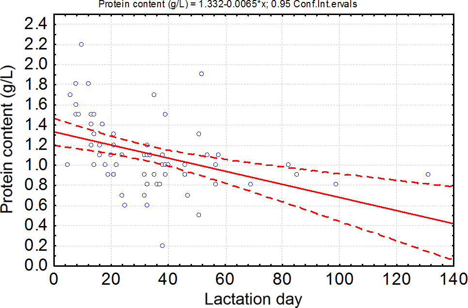
Kreissl A, et al. Human Milk Analyser shows that the lactation period affects protein levels in preterm breastmilk. Acta Paediatr. 2016;105(6):635-40. Preterm breast milk protein content soon after initiating lactation is higher than later on. This is new data confirming that, and graphically showing how enormously variable the protein concentrations are. One was only 0.2 g/L

breast milk protein

Protein concentration of milk expressed by mothers of preterm infants during the lactation period. We included 70 milk samples expressed by mothers of preterm infant between the 5th and the 131st day of lactation in the analysis.

This is one of the things making enteral nutrition with maternal breast milk difficult. Breast milk fed babies, as a group, grow more slowly than formula fed babies, often because some of them aren’t getting enough protein. As yet, breast milk fortification is standardized, but these types of variability really support the idea that individualized fortification might be preferable. Right now the difficulties with frequent breast milk analysis mean that a baby receiving breast milk and not growing adequately gets a blind increase in either feeding volume or fortifier; whereas they might benefit more from just a protein supplement for example.

Einloft PR, et al. Supplemented vs. unsupplemented human milk on bone mineralization in very low birth weight preterm infants: a randomized clinical trial. Osteoporosis International. 2015;26(9):2265-71. This trial in VLBW infants confirms that human milk fortifiers improve bone mineralization when they receive human milk. There is nowhere near enough phosphorus and not enough calcium in human milk for very preterm infants.

Omarsdottir S, et al. Cytomegalovirus infection and neonatal outcome in extremely preterm infants after freezing of maternal milk. Pediatr Infect Dis J. 2015;34(5):482-9. In a previous NICU we decided to routinely freeze all breast milk for preterm infants, because of lab evidence of CMV inactivation during freezing and thawing. In this RCT 140 infants were randomized to either have their mother’s milk frozen before it was fed to them, or to receive unfrozen milk. There was no overall difference in CMV transmission, many of the mothers produced CMV positive milk. When the analysis was restricted to those with CMV positive milk, the CMV transmission rate with CMV-DNA positive milk was 8% (3 of 37) in the intervention group and 6% (2 of 33) in controls. Freezing the breast milk reduced the rate of serious fungal infections however, which is possibly a true effect of the freezing, or perhaps a spurious result as they did make a large number of comparisons.

So let’s go back to the study from Corpeleijn,  A secondary analysis of their data, that is, in essence, an observational study using their data showed that the infants who received more than 50% of their feeds as mother’s milk (which was supplied non-pasteurized) had many fewer infants with the primary outcome. Breatmilk

The groups were diverging before 10 days of life.

It may be that unpasteurized maternal milk is the best source of milk for very preterm infants, with fewer infectious/NEC complications. But there will never be an RCT of maternal vs donor milk so we’ll just have to accept that, I think. The benefits of donor milk aren’t as clear, but there may be a reduction of NEC compared to the rates with formula, a reduction which isn’t evident if the comparison is just for 10 days. Human milk needs fortification to optimize bone mineralization, and composition is so variable that often extra fortification will be needed to get to optimal protein intakes. CMV transmission is very frequent and doesn’t seem to be prevented by freezing the milk; the importance of postnatal CMV transmission is something that has been studied recently in several investigations… there could well be a post about that!

Posted in Neonatal Research | Tagged , , , , | 6 Comments

The multidisciplinary conference, a parental view.

When patients are more complex than average, or there are multiple complications, or difficult ethical issues, we frequently organize “multidisciplinary conferences” which involve all the medical consultants and nursing, often also our social work and/or psychology personnel and the spiritual care person, sometimes respiratory therapy and other therapists as well. We may do this to try to hash out difficult issues, make sure we are all on the same page, to make plans for care and to make sure that we have discussed the different possible approaches among ourselves. Most of our conferences don’t currently include the parents, but the parents are told the conference is going on, and someone will talk to them about it afterward.

The reasons for not including parents have been multiple, and vary from patient to patient, sometimes the issues are very technical, sometimes feelings in the team are running high, sometimes we are really not sure what to do and want to discuss the limits of what is acceptable.

I’ve often thought it would be better to have parents at many of our conferences, I think that, apart from some of the more technical issues, we should have parents as part of the multidisciplinary group; on the other hand there are often strong disagreements (sometimes even arguments) and I wonder if it would help parents to be present, or might harm them.

What I mean about the technical issues is illustrated by a hypothetical case of a child with pulmonary atresia and oesophageal atresia. The baby needs interventions for the heart and for the oesophagus, but is stable on prostaglandins. We have to plan when each of these is going to be done, in what order, and what to do about the other issue while one issue is being addressed. I think that a discussion like that could be with or without the parents, based on their wish to be there or not. On the other hand if that same baby was also very preterm then the questions being asked will also include a discussion of whether ongoing intensive care with multiple procedures and a reduced chance of long-term survival is appropriate and has as much to do with values as it does with the very limited statistics about survival that might help. In that circumstance, surely the parents should be there (unless they don’t want to be).

One thing I don’t think we should aim to do when the questions are about ethics and limits of care and so on, is to finish the conference with a single plan. To come out of the conference with a decision that palliative care will be instituted, for example, would be completely inappropriate. That is a decision that the parents must be involved in.

I think health care professionals often think that it is important that we all present the same message, that there is consistency and consensus and that parents are not exposed to the differences of opinion that are the reality of all human decisions. But how do parents think about that?

The view point of the parents is something that has rarely been heard, however. One very strong opinion was recently posted over at Rumer’s Rainbow, a blog  from the parents of a child with trisomy 18. The title of the post says a lot  “The “professionals’ meeting”: creating a consistent message or colluding against parents?”

I’d like to quote a few paragraphs from the post, and invite you all to go and read the whole parental perspective.

The professionals gather in a room, discuss the child’s case and make a plan of action. Sometimes they feed back to the parents, sometimes they don’t. The reason given for these meetings is so that professionals are able to adopt a consistent line, as parents report in research and in real life that inconsistency between health professionals worries them, upsets them or angers them.

I don’t disagree that consistency matters. It is very difficult when one day you are told your child is doing really well, and the next day, someone else tells you that actually they’re really concerned. Or one day a doctor tells you an x-ray is fine and the next day another doctor tells you the same x-ray is uninterpretable, or…actually I could give you about a thousand examples from Rumer’s life.

However, I think the problem isn’t so much with inconsistency; it is a problem with honesty. A problem with saying “In my opinion…”; a problem with acknowledging disagreement: “I think X but another doctor thinks Y”; a problem with not wanting to be the bearer of bad news: “Actually your child is not doing as well as we hoped”; a problem with arrogance and pride when they say “X is the best” rather than “My view is that X is the best”.

Parents can cope with honest inconsistency – after all, I think most of us know that medical professionals are fallible humans, and we also know that our child is complex.

Rumer’s parent goes on to imagine what it would be like in “A hospital which truly values collaborative working with parents and does not infantilise them, does not see them as one with the child but as one with the professionals, part of the care team”.

The blog has lots of other good stuff on it. The most recent post for example, a letter written to a major hospital in the UK, or a post which might make you uncomfortable called “How to counsel parents: A guide for consultant neonatologists, part 1“. Personally I am waiting for parts 2, 3 and 4.

Posted in Neonatal Research | Tagged , | Leave a comment

Erythropoietin for Asphyxia

A preliminary RCT in babies undergoing therapeutic hypothermia (Wu YW, et al. High-dose Erythropoietin and Hypothermia for Hypoxic-Ischemic Encephalopathy: A Phase II Trial. Pediatrics. 2016). They randomized 50 babies with HIE who were being cooled to 1000 units/kg on days 1,2,3,5, and 7, or placebo. The primary outcome is reported as being 1 year outcomes.

The registration document says something quite different. The registration record states that the primary outcome is “Markers of organ function- The investigators will monitor organ function and adverse events until hospital discharge from the neonatal intensive care unit”. That record also states that the secondary outcome is the Alberta Infant Motor Scale, and the “other outcomes” include the Warner Initial Developmental Evaluation of Adaptive and Functional Skills (WIDEA) which is a questionnaire filled in by parents about a young child’s functional outcomes.

The published article, in contrast, states that the primary outcome was “12 month neurodevelopmental outcome assessed by (1) AIMS and (2) the Warner Initial Developmental Evaluation (WIDEA), The WIDEA was also administered at 6 months of age. Moderate to severe neurodevelopmental impairment at 12 months was defined as an AIMS score less than the fifth percentile for age, or a WIDEA score >2 SDs below the mean based on normative data from typically developing infants at 12.9 months of age.”

I was very confused by this part of the document for a while, but I think the phrase “The WIDEA was also administered at 6 months of age” is just in the wrong place, and should have been put later in that paragraph. They don’t explain why they used 12.9 months, and a threshold of 76.4, for the WIDEA test. I don’t know anything about that test, and can’t find a lot of information on-line.

I also don’t know if the planned analysis was a comparison of the mean scores, or the proportion of babies with the composite “moderate to severe neurodevelopmental impairment”.

The registration document also says nothing about MRI.

There were 42 babies who survived and were seen in follow up; there were some differences in motor scores, both the mobility part of the WIDEA and the AIMS,  which were improved in the group that received erythropoietin. But there was no difference in the proportion who were classified as “moderate to severe”. There were also some differences in the MRI brain injury scores, showing less cerebellar and subcortical abnormalities.

Encouraging, but by no means definitive, we need more data before this becomes routine. Fortunately there are some in progress, so we  shouldn’t have to wait too long. Also good news is that it seems to be a very low toxicity intervention, even in this group of intensive care dependent babies. Once we are sure of efficacy the only thing to stop its use would be the cost, which is not huge.

This study also points out the importance of registration of trials. Trial registration was initially conceived of as a way to reduce publication bias, it is essential to register trials before they are started, and to ensure that the eligibility criteria and outcomes are accurately recorded. If there is a good reason for changing those criteria or outcomes after registration, this should be explained in the text of the publication. Many trials are published and the outcomes don’t match, which can raise all sorts of questions. Trial registration is not just an extra chore for investigators, but an important part of the performance of a clinical trial, and is, in fact, one of the less onerous parts. It doesn’t take a lot of effort to get an accurate registration document in place.

One of the things that the Cochrane Collaboration has introduced into the “risk of bias” evaluation is an evaluation of whether the reported outcomes match the planned outcomes, for which the registration documents, or a published protocol if it is available, are used. For older trials it is often impossible to find the information, but more recent, registered trials often show some differences between the planned and the reported outcomes, which is rarely well explained.

Posted in Neonatal Research | Tagged , , | Leave a comment

Necrotizing Enterocolitis: manipulating the microbiome, part 3

Abdulkadir B, et al. Stool bacterial load in preterm infants with necrotising enterocolitis. Early Hum Dev. 2016;95:1-2.  More work from the Newcastle group, this time the group decided not to look at the proportion of different bugs in the stools, but the total bacterial copy count number. They took stools from 10 babies with NEC and 10 controls. When the total bacterial load was analyzed, there were no significant differences.

Dutta S, et al. Comparison of stool colonization in premature infants by three dose regimes of a probiotic combination: a randomized controlled trial. American journal of perinatology. 2015;32(8):733-40. This RCT compared 4 groups (n=149 total), no probiotics in one group, and 3 different doses in other groups. Their preparation included Lactobacillus acidophilus, Lactobacillus rhamnosus, Bifidobacterium longum, and Saccharomyces boulardii. They received either a billion a day for 21 days, 10 billion a day for 21 days, or 10 billion a day for 14 days. The main outcome they were investigating was the rate of stool colonization. Almost none of the babies grew Saccharomyces, about half of the controls and over 80% of the treated babies grew Lactobacilli, around about 1/3 of controls, and 80% of treated babies grew Bifidobacteria. There were no real differences between the treated groups. They also did quantitative cultures, and the median probiotic bacterial counts for all species in control babies was zero, and were very much higher in treated babies. The counts tended to be a bit higher with the highest dose group, but these data suggest that the dose of the probiotic regime may not be very important: as they are live organisms and we are hoping for colonization and growth in the intestines, I don’t find that very surprising.

Kanic Z, et al. Influence of a combination of probiotics on bacterial infections in very low birthweight newborns. Wien Klin Wochenschr. 2015;127 Suppl 5(5):S210-5. This was a small RCT (n=80) which was not included in the recent systematic review of probiotics to prevent invasive late-onset sepsis in very preterm infants that I discussed recently. A mixture of Lactobacillus acidophilus, Enterococcus faecium and Bifidobacterium infantum  substantially decreased the rate of sepsis, which is consistent with the results of the systematic review.

And now some Necrotising Enterocolits research that isn’t specifically about the microbiome.

Shin SH, et al. Surgical Necrotizing Enterocolitis versus Spontaneous Intestinal Perforation in White Matter Injury on Brain Magnetic Resonance Imaging. Neonatology. 2016;110(2):148-54. 9 very preterm babies with spontaneous perforation were compared to 24 with surgical NEC. The white matter injury score was significantly worse with surgical NEC.

Schat TE, et al. The relation between splanchnic ischaemia and intestinal damage in necrotising enterocolitis. Archives of disease in childhood Fetal and neonatal edition. 2016. The authors followed 19 preterm babies with NEC during 48 hours after onset of the condition, 10 of whom had surgery or died (complicated NEC), and 9 did not (uncomplicated). Using NIRS to investigate intestinal oxygenation:

IFABPp

The uncomplicated NEC cases seem to have had hyperperfusion at the start, with low oxygen extraction, which gradually got back towards normal. The complicated cases had relatively high values at first, maybe because their perfusion was limited, and this progressively fell, perhaps due to necrosis. The I-FABPp levels (Intestinal Fatty Acid Binding Protein in plasma) an indicator of disease activity in NEC, were higher in the complicated cases.

Shah SD, et al. Early versus Delayed Human Milk Fortification in Very Low Birth Weight Infants-A Randomized Controlled Trial. The Journal of pediatrics. 2016. This is an RCT in 100 VLBW infants, who were receiving either maternal or donor breast milk. They were randomized to have breast milk fortifier (Mead Johnson Liquid Fortifier, supplied by the manufacturers who also funded the trial, but didn’t have much to do with the trial otherwise, they didn’t design or analyze it), started at 20 mL/kg/d of feeds, or to wait until they reached 100 mL/kg/d. The study doesn’t have a lot of power for safety outcomes, but it is I think the first trial to examine this issue.

The feeding protocols are painfully slow, babies under 800 g for example “received trophic feedings (10 mL/kg/d) for 3 days and advanced by 10 mL/kg/d every other day”. But at least they were the same between the two groups.

There were no differences in days to full feeds (that is, the early fortified feeds were tolerated as well as the late fortified feeds) or in NEC, or in other adverse outcomes. The only significant difference is that the early fortified babies received more protein over the first 4 weeks of life. Somehow the babies received the same calorie intake over these 4 weeks, which I guess must be because this was an unblinded study, and the doctors decreased the intravenous calories when the babies got early fortification, to match the calorie intake of the late fortified. But they didn’t do the same thing with intravenous protein.

Despite getting more protein, they still didn’t get enough, (at least according to the ESPGHAN standards which recommend 4 to 4.5 g/kg/d) they averaged around 3.5 g/kg/d; and they didn’t get enough calories either, averaging 110 kcal/kg/d when on full feeds, whereas 120 or more is preferable.

Growth (in weight) was a little better in the early fortified group, but they still lost 1.3 standard deviation of weight between birth and 36 weeks.

In order to get better growth outcomes, more protein, and more calories, and an on-going emphasis on adequate nutrition throughout the NICU stay are required.

Early fortification is probably safe, according to this trial, so integrating it into a comprehensive approach to improve nutritional outcomes is appropriate.

As I have noted before on this blog, there are no reliable data that show that how we feed babies has any influence on NEC, when we start, how we advance, when we advance etc. none of those things have been shown to affect the incidence of Necrotising Enterocolitis. The only exception to that general rule is a single trial of 140 preterm infants that kept the babies nil by mouth for an average of 10 days after birth, and then randomized them to get 10 days of small volume feeds compared to immediate feed advancement. That trial was stopped early after 1 small volume baby and 7 of the immediate advancement babies developed NEC. So if you keep your babies without feeds for too long, it might make a difference if you use a period of “trophic feeding”. (one of their NEC babies had been npo for 31 days before the intervention started!) Otherwise there is no evidence from any other intervention trial that feeding patterns change whether a baby develops NEC or not. Despite this there have been many publications of groups that state something like “we have low NEC rates because we hardly feed our babies any milk”. I think that much of the difference in NEC rates between NICUs is likely due to differences in the bacteriology of different NICUs, and different acquisition of the intestinal microbiome between those units. Something that we can manipulate, with some degree of success.

Posted in Neonatal Research | Tagged , , , , | 2 Comments

Necrotizing Enterocolitis: manipulating the microbiome, part 2

The Newcastle, UK, group has published an article about the intestinal microbiome of babies receiving routine probiotic prophylaxis. Abdulkadir B, et al. Routine Use of Probiotics in Preterm Infants: Longitudinal Impact on the Microbiome and Metabolome. Neonatology. 2016;109(4):239-47. The authors studied 7 extremely premature babies who received Infloran (Bifidobaterium bifidum, and Lactobacillus acidophilus) and 3 controls, also extremely premature.

Basically, they showed greater Bifidobacterium (15.1%) and Lactobacillus (4.2%) during supplementation compared to the control group (4.0% and 0%, respectively). Bifidobacterium counts stayed high following discharge, but the Lactobacillus disappeared.

Several previous studies in very preterm infants have shown extremely low, or absent, Bifidobacterial colonies, increasing that to 15% shows an enormous change in the intestinal microbiome.

The impacts of the microbiome changes on the “metabolome” of gut are also presented, the results seem to me preliminary, and as yet of uncertain significance, but there appear to be some differences between babies getting probiotics and those not.

Some of the same authors have written a review article Embleton ND, et al. Probiotics for prevention of necrotizing enterocolitis and sepsis in preterm infants. Curr Opin Infect Dis. 2016;29(3):256-61.  It is fairly brief and summarizes the data well, the abstract ends like this:

Despite current uncertainties, it is difficult for clinicians to ignore the current data, and increasing numbers now use commercially available products. It remains a matter of concern that many products lack the robust quality control most clinicians and parents would consider important for use in vulnerable populations. Head-to-head trials are needed.

Which I think is right on the money. And also brings me neatly to the next article:

Lewis ZT, et al. Validating bifidobacterial species and subspecies identity in commercial probiotic products. Pediatr Res. 2016;79(3):445-52. Mark Underwood’s group from UCDavis examined the composition of 16 formulations of probiotics that were promoted as containing Bifidobacteria, some available on-line, and others in local stores. One of the complicating factors in this field is the changes in classification that occur, so the same strain can go from being an B. longum to being a B. animalis.

They found that the advertised content of many of the products varied significantly from the true composition. Only 1 of the 16 products tested had exactly the same Bifidobacteria species in every sample as what was written on the label. In addition:

Some products were not internally consistent as both pill-to-pill and lot-to-lot variation were observed

Some producers in other parts of the world have better quality control, and more reliable composition, but it is really hard to know,if you just go to the local health food store, if you are getting what you think.

When we started using probiotics routinely a few years ago, we were fortunate that there were preparations with good quality control, and which followed GMP. Standards for probiotics for premies are extremely important.

Mark Underwood is also an author of a recent review article, Vongbhavit K, Underwood MA. Prevention of Necrotizing Enterocolitis Through Manipulation of the Intestinal Microbiota of the Premature Infant. Clin Ther. 2016;38(4):716-32. Another excellent review, the last sentence in their abstract is the following :

Shaping the intestinal microbiota of the premature infant through human milk feeding and dietary supplements decreases the risk of NEC. Further studies to identify the ideal microbial composition and the most effective combination of supplements are indicated.

Posted in Neonatal Research | Tagged , | 2 Comments

Necrotizing Enterocolitis: manipulating the microbiome, part 1

Some very interesting recent publications about NEC, so I will post a multi-part bumper posting.

The first has just been published (in tomorrow’s Lancet) Warner BB, et al. Gut bacteria dysbiosis and necrotising enterocolitis in very low birthweight infants: a prospective case-control study. The Lancet. 2016;387(10031):1928-36.

In this study the authors collected stool samples from all the VLBW infants in their NICU and froze them. They started doing this in St Louis, and then added cohorts from two other NICUs. When a baby developed NEC they analyzed all the stools up to the day of diagnosis (max 60 days), and then they found some matched controls (1 to 4 per case), with similar gestational age and birth date, they analyzed all their stools up to 30 days of age, and then every 3rd day to 60 days of age. What they call the “primary cohort” are the babies from St Louis, the “secondary cohort” was the babies from the other 2 centres, where stool collection started later.

In total they analyzed 2492 stool samples from 122 infants in the primary cohort, of whom 28 developed NEC; there were 94 controls.

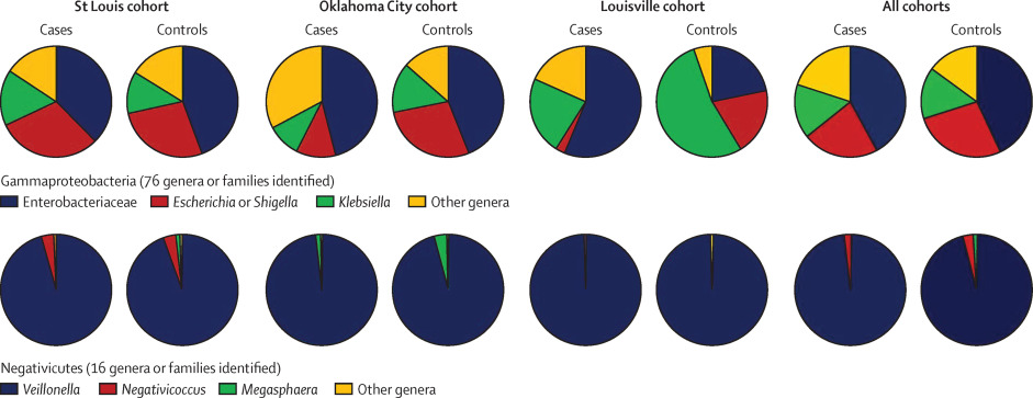
The microbial community structure in case stools differed significantly from those in control stools. These differences emerged only after the first month of age. In mixed models, the time-by-necrotising-enterocolitis interaction was positively associated with Gammaproteobacteria (p=0·0010) and negatively associated with strictly anaerobic bacteria, especially Negativicutes (p=0·0019).

In the secondary cohort there were 44 infants, of whom 18 developed NEC. The results from the combined cohorts reflected what that quote above notes, with the additional finding that there was an decreased proportion of the combined Clostridia–Negativicutes class (p=0·0051) in the NEC babies compared with controls.

I am no microbiologist, so the term “Negativicutes” was new to me. It refers to a class of the Firmicutes that are Gram negative, (most Firmicutes are Gram positive, including clostridia and bacilli).

The Gammaproteobacteria include lots of the bad boys, Pseudomonads, and the Enterobacteria, which includes Salmonella, E Coli, Enterobacter, and Klebsiella.

Cuties

This simplified figure in the article points out some of the complexity of the situation, if you compare the controls between centres you can see marked differences, for example in the proportion of the Gammas that are Klebsiellae, much higher in Louisville than elsewhere. I think this actually may account for some of the differences in NEC rates between centers, the microbiology of your NICU could have a major influence on NEC, much more than how you feed the babies!

In addition, the article confirms other observations that the intestinal microbiome of babies that go on to develop NEC is much less diverse than controls.

As the accompanying editorial notes the “relative abundance of Gram-negative facultative bacilli and relative paucity of anaerobic bacteria (ie, Clostridia and Negativicutes) could represent the microbiota signature preceding necrotising enterocolitis in very low birthweight infants.”

bifidos

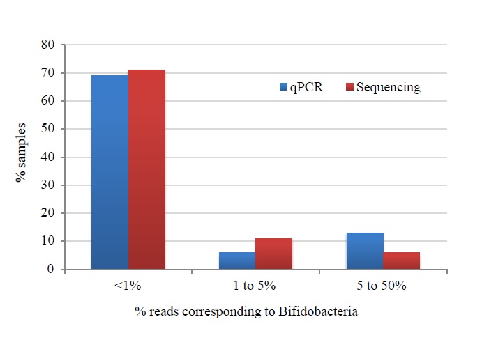
The authors, Warner et al. also used other techniques to examine Bifidobacteria specifically, and as this figure shows (which is from their on-line appendix), using 2 different methods (the red and the blue bars), there were very few Bifidobacteria in their babies, most having <1%. There was also no difference between cases and controls. They state the following:

 Our data also provide a microbiological explanation for the recent failure of the probiotic Bifidobacterium breve BBG-001 to prevent necrotising enterocolitis in a large and rigorous multicentre randomised controlled trial in the UK. Specifically, we found no evidence that Bifidobacteria were under-represented in the stools of infants who subsequently developed necrotising enterocolitis.

Which is true as far as it goes, but there were vanishingly few bifidobacteria in either group. The reason for the negative results from the Pips trial do need to be figured out, it may be the wrong strain, or just randomly lower effect than the other trials, with whom the 95% confidence intervals overlap. On the other hand, the Newcastle group have recently published data showing very much higher bifidobacterial concentrations among infants who received routine probiotics compared to non-treated babies.

Which I will discuss in part 2.


Posted in Neonatal Research | Tagged , | Leave a comment

Which Surfactant is best?

Singh N, et al. Comparison of animal-derived surfactants for the prevention and treatment of respiratory distress syndrome in preterm infants. Cochrane database of systematic reviews (Online). 2015;12:CD010249.

This is the sort of systematic review that I find really helpful. Some (including some of my own) end up finding little good evidence, and summarizing the results of only one or 2 small trials. That may help to point out the lack of evidence, and to stimulate future trials, but doesn’t often give enough information to really inform practice, and individual treatment decisions.

In contrast, this review includes a significant number of trials, some with respectable sample sizes, and ends up  finding significant differences between some of the surfactants that are available.

The review divides the surfactants into the following:

bovine lung lavage surfactant extract, which includes Infasurf, bLES (the Canadian one) and Alveofact

bovine minced lung surfactant extract, which includes Survanta and Surfacten (the old surfactant TA, the Japanese one)

porcine minced lung surfactant extract, which I  know as Curosurf,

porcine lung lavage surfactant, which is apparently called Surfacen.


The authors searched for all the comparisons that had been studied.

Most of the comparisons showed no difference in important clinical outcomes, (although some comparisons had relatively little power,) for example, bovine minced lung and bovine lung lavage surfactants were basically equivalent for all the outcomes. This was true both for prophylactic use and for rescue use.

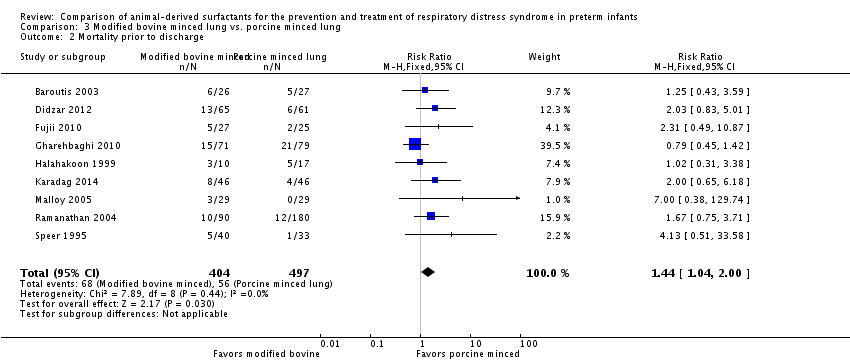
The comparison that did show different effects was the comparison between minced bovine lung, and minced porcine lung (that is between Survanta and Curosurf), these were all rescue/treatment trials: The first outcome was mortality.

surf death

As you can see the studies are all fairly small, and individually do not show a difference, that is always a bit of a worry, as multiple small studies can inflate an effect. Nevertheless, the summary effect shows higher mortality with the bovine minced lung preparations than the porcine surfactants.

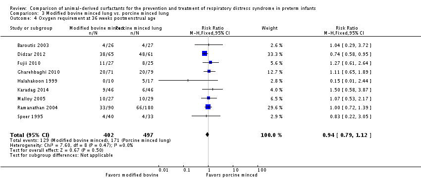
The next outcome is ‘BPD’ or oxygen requirement at 36 weeks.

surf bpd

Here there is no sign of a difference, when the outcome is expressed as a proportion of those who were enrolled into the study.

The authors of the review also present the outcome “death or BPD” which, as those of you who were at my talk in Baltimore know, I think is not an appropriate composite outcome, and those who were at the International Neonatal Collaboration INC meeting in Bethesda recently know that many of the leaders in the field are of the same opinion. The outcomes of death and BPD have different value for parents (and for society as a whole) and may not change in the same direction. I think the outcomes of Death as one outcome, and BPD as a separate outcome among surviving infants, make much more sense. Unfortunately you can’t always tell from data as presented, but, in this case, if we make the probably unsafe, but probably not very wrong, assumption that all the deaths were before 36 weeks, then you can estimate the BPD among survivors. You come up with a relative risk of exactly 1.0, (95% CI 0.85, 1.18).

So overall it seems that porcine minced surfactant leads to more survivors, but the same proportion of BPD (that is O2 requirement at 36 weeks) among surviving infants.

As I mentioned, the confidence intervals are wide, and there are several small studies, but despite these concerns, there are relatively fewer deaths with porcine surfactant, with a risk difference of 0.05 and an NNT, for one fewer death, of 20. These data suggest therefore, that if you treat 20 babies with poractant rather than a minced bovine lung preparation, there would be one more survivor.

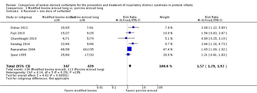
Also there are substantially fewer babies who need a second treatment.

surf dose

The NNT here is 7, which means that for every 7 babies you treat, one extra baby will need a second dose. (In the babies in the review, a second dose was required for about 40% of the bovine and about 25% of the porcine treated).

Pneumothoraces were a bit less frequent with porcine, maybe due to chance, and if all airleak syndromes were put together, there were fewer airleaks with porcine.

For the other comparison which is of specific interest to me, that is minced bovine lung to bovine lung lavage surfactants, almost all of the data comes from the trials of Bloom which compared calfactant (Infasurf) to beractant (Survanta). Neither the prophylactic nor rescue trials showed any differences.

The manufacturers will I am sure moan that not all the surfactants within each of these groups are identical, and indeed there are some differences in the details. Calfactant, for example has around twice as much Surfactant Protein B and C compared to bLES. But there is almost no comparative data available for bLES.

Why is the porcine preparation better, that is it works faster, leads to fewer airleaks and has lower mortality?

The studies generally used the manufacturers recommended dose, which is higher for poractant than for the bovine preparations. That may be the only reason, I am not sure about that, but in any case poractant alpha is concentrated enough that the higher initial dose is easily tolerated, whereas it would mean quite large fluid volumes for the other preparations.

In Canada we are still waiting for Curosurf. It has been approved, over a year ago, but is still not being marketed. Come on Chiesi! We are waiting…

Posted in Neonatal Research | Tagged , , , , | 2 Comments