How fast to feed?

One of the recurring themes in this blog is that good, large enough, prospective RCTs do not necessarily reproduce the results of prior smaller trials, and often do not reproduce the findings of observational studies. Specifically, I have mentioned previously that observational studies suggesting that slow feeding advancement leads to less Enterocolitis in extremely preterm babies (NEC) than faster advancement have never been confirmed by prospective trials.

There were several trials of feeding advancement, of variable quality and relatively small sample sizes, before 2016, which showed no overall adverse impact of advancing feeds more quickly, but with sufficient uncertainty that a large simple pragmatic trial was warranted to address the question. The short term results of the SIFT trial have been presented and incorporated into the Cochrane review Oddie SJ, et al. Slow advancement of enteral feed volumes to prevent necrotising enterocolitis in very low birth weight infants. Cochrane database of systematic reviews (Online). 2017;8:CD001241.

Those results showed no benefit of advancing feeds more slowly (18 mL/kg/day) compared to more quickly (30 mL/kg/day) among nearly 3000 infants of less than 32 weeks, or less than 1500g birth weight. The findings incorporated into the Cochrane review include secondary outcome data on NEC and infections.

The primary outcome of the SIFT trial was survival without neurodevelopmental disability to 24 months corrected age.  In the latest FPNEJM the primary and the secondary short term outcomes are now presented in full. Dorling J, et al. Controlled Trial of Two Incremental Milk-Feeding Rates in Preterm Infants. New Engl J Med 2019;381(15):1434-43. 2800 infants were randomized when receiving less than 30 mL/kg/day and the intervention started when ‘the clinicians were ready to start increasing feeds’ which occurred at a median of 4 days of age. The more rapid increase group reached ‘full feeds’ which was defined as 3 days of receiving at least 145 mL/kg/d, after a median of 7 days, compared to 10 days in the slower group. I don’t understand this entirely; if the baby was on 10 mL/kg/d and tolerated all the feed increments, it would take 5 days to get to at least 145 mL/kg/d and then another 3 days would be needed before satisfying full feeds criteria, which takes us to 8 days minimum. Anyhow, this is a minor quibble, on average the babies in the slow advancement group  had 2 days longer with intravenous nutrition. I can’t see in the publication any definition of feeding intolerance, or if there were any attempted standardization of feeding approaches to decide whether to continue advancing feeds.I beleieve the investigators decided to leave most details of feeding approaches untouched, only mandating the attempted feeding volume changes, and counting on a very large sample size even out everything else.

NEC was very slightly less frequent with faster feeds (5% vs 5.8%, RR=0.90, compatability intervals 0.66, 1.24) and late onset sepsis or clinical sepsis were very slightly less frequent also (30 vs 31%, RR=0.96, compatability intervals 0.85 to 1.08).

The differences between groups in the primary outcome also were tiny, survival was 95% in each group and so-called disability was 31% fast, vs 28% slow. Survival without ‘disability’ was 66% for the fast feeders and 68% for the slow. I won’t go into the details of how the outcome assessments were performed, most were by parent report, others by direct observation and evaluation.

In subgroup analyses published in the on-line supplement, there is no clear evidence of a difference in NEC or of nosocomial infections by gestational age subgroup. Infants with reversed or absent end-diastolic flow on antenatal doppler ultrasound of the umbilical artery also had no clear difference (although in this subgroup faster feeding advancement had rather less NEC, 3.8 vs 7.1%, this may have been due to chance effects in this subgroup (435 babies) and the interaction term was compatible with a chance effect).

In the Cochrane review mentioned above data on ‘invasive infections’ are presented, which refers to culture-positive infections. The number of infants with at least one invasive infection was 247/1389 vs 267/1397 in the SIFT data included in that review (I can’t find those data in the new publication) that gives a relative risk, favoring faster feeds, of 1.07 (compatibility limits 0.92, 1.96) when the data from the other 600 babies included in all the other trials is added, the RR is 1.15 (95% limits 1.00, 1.32). Similarly the data from all trials in the meta-analysis, including SIFT data, show a slightly higher rate of NEC with slow feeding RR=1.07, 95% compatability intervals 0.83, 1.39).

SIFT is consistent with the previous trials, but gives enormously more precision to the estimates, it shows that feeding faster, up to a goal of 30 mL/kg/ has no adverse impact overall, or in any subgroup, compared to feeding slower (goal of 18 mL/kg/d). Even though the babies had a median of 2 days less of parenteral nutrition that did not lead to a clear reduction in proven and suspected infections, there seems to be a small reduction if all the available RCT data are analyzed.

Of note, the late-onset sepsis rates appear to be extremely high; 540 of the 889 babies under 28 weeks had a suspected or proven infection, or 61%. But of course that includes ‘suspected infection’ which basically means anyone with any sort of clinical deterioration that is treated with antibiotics for more than 48 hours. If I compare the total numbers of proven and suspected infections in the final publication (848) to the culture positive infections in the Cochrane review (514), then about 60% of their LOS is culture positive. If the same proportions hold in smaller gestation age groups, then about 36% of babies under 28 weeks had a culture positive sepsis. That approaches the kind of incidence of culture positive infections in other multicenter databases, but remains somewhat high. In the 2017 CNN report, for example, adding together the <25 weeks and the 25 and 26 week gestation babies ((i.e. the higher risk infants, excluding those at 27 completed weeks) the proportion with at least one infection, culture positive, is 28%.

The trial was powered to have a reasonable chance of showing a difference in sepsis, based on the assumption that 1000 fewer catheter-person days per 250 infants would lead to fewer infections. In fact there were about 500 fewer parenteral nutrition days (I can’t find the data for catheter-person days) per 250 enrolled infants, or about 5000 fewer days overall, which didn’t seem to have any measurable effect on infections. Even when looking at culture positive infections alone, there was really nothing there. There are a number of potential explanations for this. As I have mentioned before, the emphasis on catheter linked sepsis is excessive in the preterm infant. Many LOS are caused by enteric gram negative pathogens, and reducing CLABSI doesn’t necessarily lead to a reduction in overall sepsis, by a logical extension, reducing catheter duration may reduce CLABSI but without reducing overall infection rates. On the other hand the CLABSI rate per 1000 patients days is lower in the first week of catheter use compared to later, and the median 2 days of increased duration of parenteral nutrition may not have exposed the babies to much difference in risk. Sanderson E, et al. Dwell time and risk of central-line-associated bloodstream infection in neonates. J Hosp Infect. 2017;97(3):267-74.

What next? I think that a trial comparing the fast feeding approach in this study could be compared with an earlier start of advancement at a slightly greater rate. The comparison approach could be to enrol babies on day 1, start feeds immediately at 2 mL/kg q2h, and start advancement immediately at say 40 mL/kg/d. In my practice we now usually start feed advancement before 4 days of age, and, if the infant is not in shock, advancement starting day 1 would be just fine by me. We would have to use temporarily a bit more donor breast milk (PDHM), which could be a downside to advancing sooner. An earlier start to advancement might lead to a greater difference in catheter and parenteral nutrition duration, and, potentially, a subsequent impact on sepsis. Perhaps we might find less CLABSI, but no less systemic infection.

So far there is no good quality prospective controlled data that any approach to starting or advancing feeds has any impact on NEC.

The evidence-based strategies to reduce NEC are to promote and support MOM (mother’s own milk), have PDHM available when MOM is insufficient, to have a standardized feeding protocol, and to use a probiotic preparation with good quality control, which contains bifidobacteria and probably a lactobacillus or a streptococcus.
Posted in Neonatal Research | Tagged , , , , | 4 Comments

Should we try?

Arnolds M, et al. Worth a Try? Describing the Experiences of Families during the Course of Care in the Neonatal Intensive Care Unit When the Prognosis is Poor. The Journal of pediatrics. 2018;196:116-22 e3.

A few times a year many of us are faced with admissions to the NICU which have marginal chances of survival. That might mean a congenital anomaly with a limited chance of survival, a complex cardiac defect without a clear surgical option but with a chance of surgical palliation, or an extremely preterm baby with a predicted <10% chance of survival.

Sometimes, after discussion with the parents, we decide to institute active intensive care; In other cases the diagnosis and the prognosis become evident after hours or days of intensive care, and we realize that there is only a small chance of the baby surviving.

It would be interesting to investigate the impact of intensive care that ‘fails’ on families. How do they hold up later? Is there a lot of “decisional regret” where families are disturbed about the intensity of the care their baby received, only to die anyway?

A group of investigators from the University of Chicago (including Bill Meadow in one of his last contributions to the neonatal literature) recently published a qualitative study examining the experiences of families with extremely high risk babies. Families from one of two tertiary NICUs were interviewed while their child was sick, and most of them again interviewed more than 6 months later, 3 families had infants who had died, and 2 others were still hospitalised.

There are many things in the article that are worth reading, and anyone trying to support families of critically ill babies could benefit from the insights provided. But I want to focus on one thing, and that is the idea of decisional regret. Health care workers often think ‘if the parents really knew what was going to happen, they would choose differently’, indeed it is one of the things which underlies the moral distress that is experienced by people working in the NICU. Prentice TM, et al. The use and misuse of moral distress in neonatology. Seminars in fetal & neonatal medicine. 2017.

In reality, decisional regret regarding neonatal intensive care decisions is quite unusual, and seems to be more frequent among those who chose not to intervene than those who chose NICU, even when it goes badly in the end.

This publication supports that interpretation, even when the outcome is poor (extremely long hospitalisation, or death, or likely disability) parents were grateful for the care their baby received, and expressed that they had no regrets about the decisions made. Even when the prospects are poor, families generally appreciate that their infants was considered to be “worth a try”. A title which I would guess was suggested by Bill as a pithy summary of the entire project!

Posted in Neonatal Research | Tagged , | 1 Comment

Why is MOM best? part 2

MOM is best, because it leads to the lowest incidence of NEC; but why? (if you didn’t read part 1, MOM is Mother’s Own Milk)

The impacts of milk processing which create the differences in milk composition, detailed in my previous post, may be the link to the increased incidence of NEC. That effect could be mediated by changes in the intestinal microbiome. There have been several recent publications about this:

Cai C, et al. Feeding practice influences gut microbiome composition in very low birth weight preterm infants and the association with oxidative stress: A prospective cohort study. Free Radic Biol Med. 2019;142:146-54.
Parra-Llorca A, et al. Preterm Gut Microbiome Depending on Feeding Type: Significance of Donor Human Milk. Front Microbiol. 2018;9:1376.
Ford SL, et al. Improved feeding tolerance and growth are linked to increased gut microbial community diversity in very-low-birth-weight infants fed mother’s own milk compared with donor breast milk. The American journal of clinical nutrition. 2019;109(4):1088-97.
Zanella A, et al. Influence of own mother’s milk and different proportions of formula on intestinal microbiota of very preterm newborns. PLoS One. 2019;14(5):e0217296.

These studies are relatively consistent, although the complexity of microbiome data and the ways in which they should be presented and analysed are still developing, and can be quite confusing to a non-specialist, like me. Overall, the fecal intestinal microbiome in MOM fed infants is more diverse, as shown by increases in alpha diversity; which are measures of how many different bacteria there are and how variable they are. Differences in beta-diversity, which are ways of comparing these diverse bacterial communities, have been found between formula, human milk and donor milk.

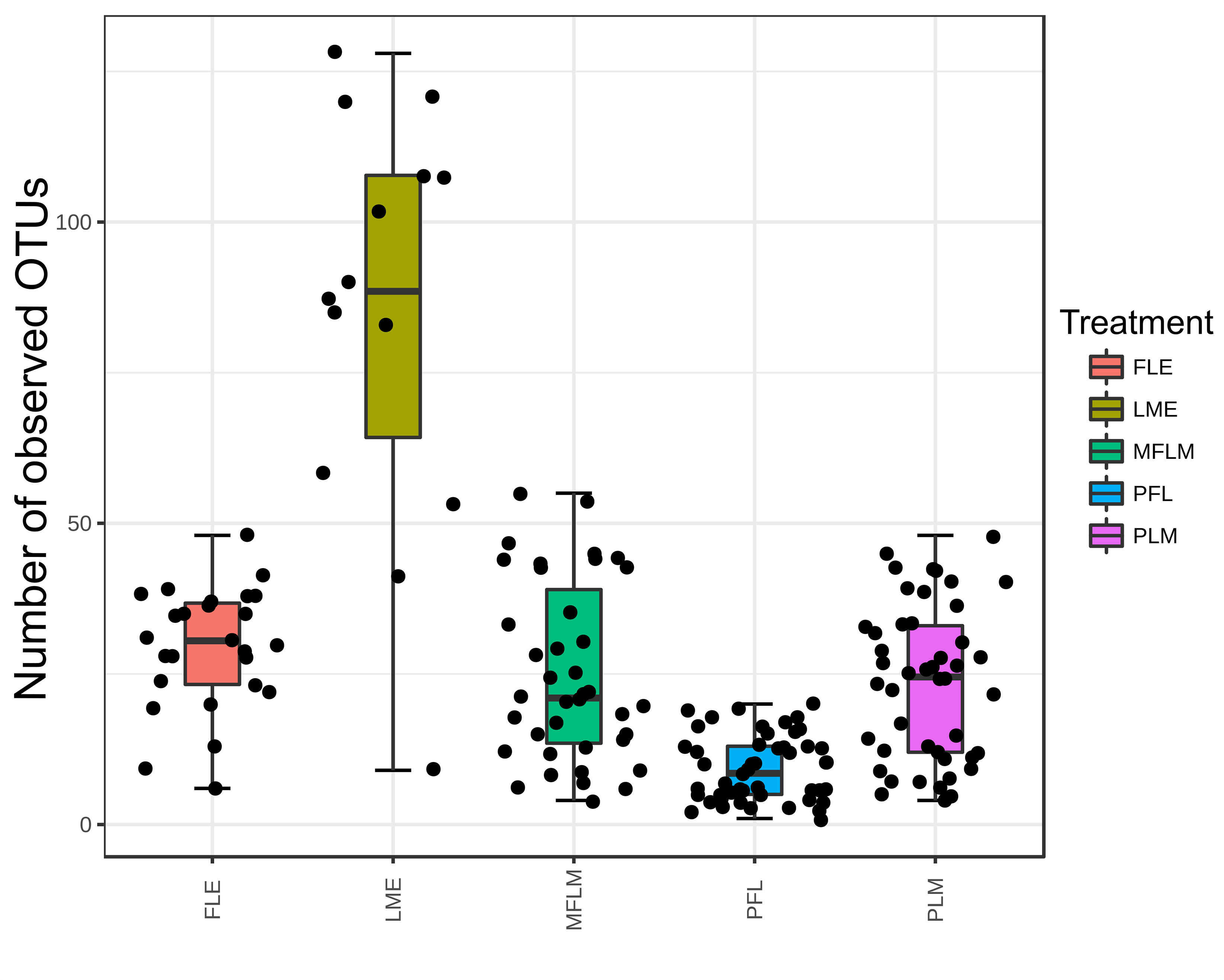
One very recent interesting study by Zanella et al reported 5 groups of babies, with exclusive MOM (LME in the figures),  exclusive formula (FLE), about 50:50 (MFLM), predominantly MOM (>70%, PLM) and predominantly formula (>70% PFL). They showed a sort of dose response, this is a box and whisker plot of the number of OTU’s.

Figure 2: Number of Operational Taxonomic Units measured in fecal samples from preterm babies fed with different diets during 28 days.

Operational Taxonomic Units is a jargon used to avoid speaking about species, as the definition of a species among bacteria is not a settled issue, microbiomologists define an OTU according to the percentage of the bacterial DNA in the 16s RNA gene which is similar to others. In this study a similarity of 97% was required to define the OTU. So there are clearly many more OTUs, and more bacterial types, with exclusive MOM compared to other types of milk feeds.

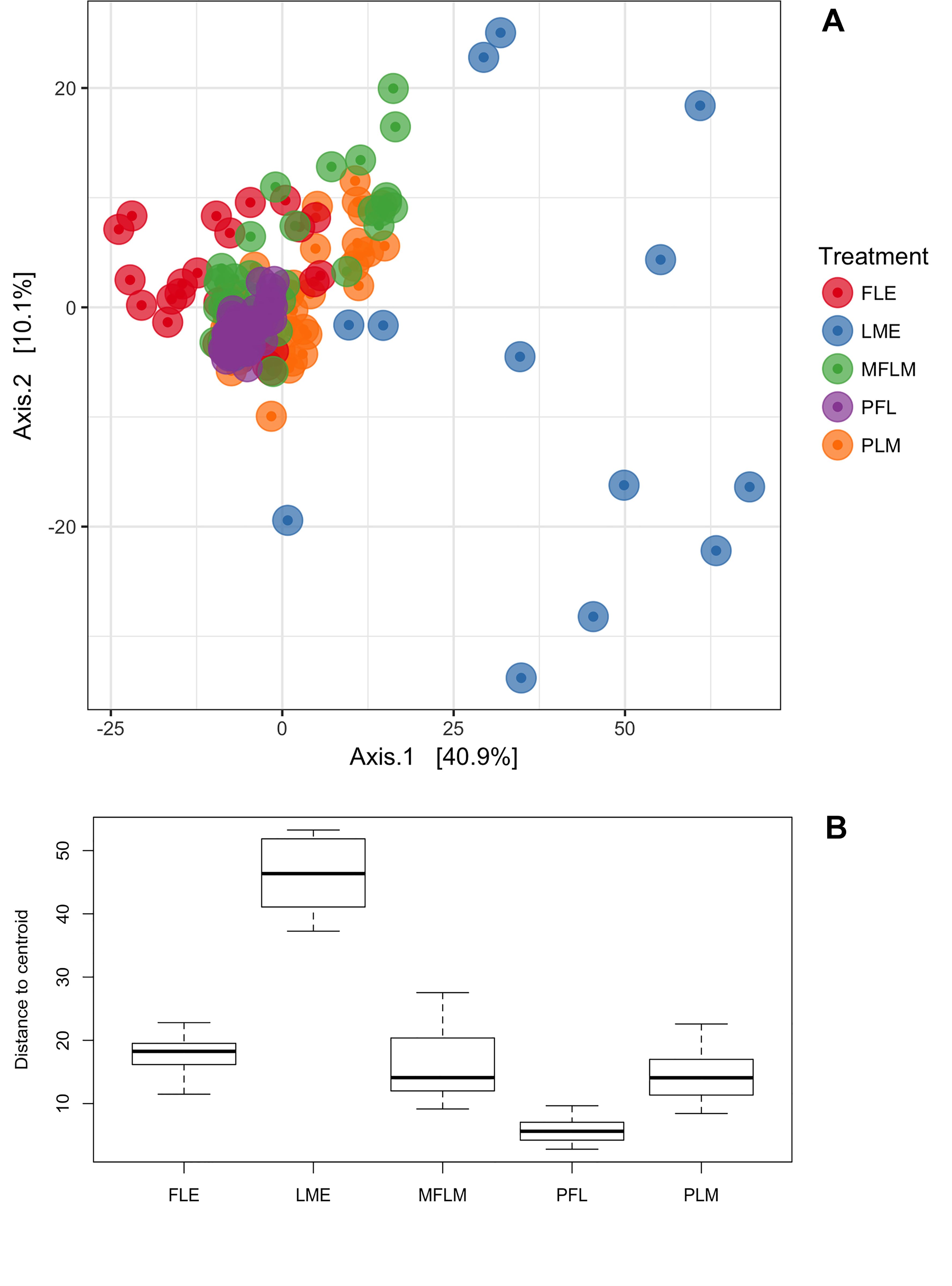
This is their graph 1, showing the beta-diversity results.

Graph A represents clusters of microbial communities. Each point represents an individual sample, with colors indicating feeding treatments. Graph B represents measurement of multivariate dispersion for each treatment.

You can see that the different feeding types tend to cluster differently, with the Exclusive MOM group (LME) the most different to the others. Interestingly, in this study the human milk was fortified with a powdered fortifier FM85 (personal communication Renato Procianoy) when the infants were at 80% of their full feeds, so the addition of the bovine protein-based fortifier does not seem to affect the microbiome diversity sustained by MOM.

Other studies looking at donor milk also show differences, with higher diversity when receiving MOM than when receiving PDHM. This might in part be because MOM usually contains bacteria classed as probiotics (particularly bifidobacteria), which are eliminated by pasteurization.

What does all this mean?

MOM is best, as usual.

Although we don’t know exactly why, MOM, which is not pasteurized or otherwise manipulated, supports a microbial intestinal community which is more diverse, and contains more bifidobacteria and lactobacillae than other milk sources. PDHM seems to have intermediate effects on NEC and on the microbiome, while formula feeding leads to major impacts on the microbiome and on the incidence of NEC.

Why does this matter?

If the adverse impacts of formula feeding are not because of the source of the protein, but due to other impacts of the processing of the milk on protein structure, milk composition, and the intestinal microbiome, then it is possible that we could produce an MOM replacement, and/or a fortification method which was able to support optimal growth without an increase in NEC. We should investigate ways of processing donor human milk which lead to less impact on intestinal function, so that when we don’t have enough MOM we can find a replacement that is just as protective.

Much more robust data on the safety of bovine fortifiers, comparison of liquid and powdered fortifiers, and proof of whether or not human milk based fortifiers are preferable, would help to maintain optimal nutrition while assuring the best outcomes for our babies.

Posted in Neonatal Research | Tagged , , | Leave a comment

Why is MOM best?

Mother’s Own Milk (MOM) seems to be the best base for enteral nutrition of the preterm infant, in terms of the risk of infection, the risk of Enterocolitis (NEC) and, probably, long term neurodevelopment.

Reasonably good data show that replacing human milk with artificial formula, when there is insufficient maternal breast milk, leads to increased intestinal inflammation and NEC, compared to using pasteurized donor human milk (PDHM).

From observational studies, (which is all we’ll ever have, as you couldn’t ethically randomise babies to a PDHM supplement if there is enough MOM), PDHM seems to be less protective than MOM.

For the baby whose mother doesn’t produce any MOM, using PDHM rather than formula seems to be better for the outcomes of NEC, and probably late-onset infecions, among very preterm infants.

But why is that the case? What is it about MOM that leads to fewer of those complications? Is it just because nature loves mothers? Is it rather that there is something about MOM which is different?

Fortifying human milk with a bovine-milk based commercial fortifier, to increase protein, calorie and mineral supply, has never been shown to increase NEC or LOS, compared to either no fortifier, or to a human milk based fortifier.

I think it is important to specify that it is very hard to prove a negative, ‘that multicomponent fortifiers do not increase the risk of NEC’ for example, and we should remain vigilant regarding the power of the individual studies and of the accumulated evidence. The latest Cochrane review states that the included studies were small and of low quality and that there is ‘low-quality evidence that fortification does not increase the risk of necrotising enterocolitis in preterm infants (typical RR 1.57, 95% CI 0.76 to 3.23; 11 studies, 882 infants)’. Which means that a 57% increase in NEC was the most likely result of using fortifiers compared to not using them, which was far from statistical significance.

Since that 2016 version of the review there is at least 1 other RCT that seems to confirm those findings (still a very small study) Adhisivam B, et al. Does fortification of pasteurized donor human milk increase the incidence of necrotizing enterocolitis among preterm neonates? A randomized controlled trial. The journal of maternal-fetal & neonatal medicine. 2018:1-6.

Also published was what is, I think, the only RCT of babies receiving MOM who were randomized to human milk-based fortifier compared to a commercial bovine milk-based fortifier. O’Connor DL, et al. Nutrient enrichment of human milk with human and bovine milk-based fortifiers for infants born weighing <1250 g: a randomized clinical trial. The American journal of clinical nutrition. 2018;108(1):108-16.  Which showed no impact of the fortifier origin.

Why is MOM best?

Because of the frequent diagnosis of cow’s milk protein intolerance in children, (much more frequently diagnosed than proven, I might add), you might suppose that an immune response to exogenous proteins is a likely answer to the increase in NEC with supplemental formula feeds. But if that were the case, why is fortifier not implicated? Also in general, exposure of preterm infants to exogenous protein leads to tolerance rather than intolerance, and other immune phenomena are quite unusual in the newborn. For example. the widespread instillation of bovine or porcine proteins into the airways of preterm infants does not seem to be related to any later adverse immune events.

If the source of the protein is not the primary problem and, I reiterate, there is no good evidence that it is, but formula still causes an increase in NEC, then the emphasis on an ‘exclusive human milk diet’ may be wrong-headed.

There are a number of clues in the literature that suggest that other factors in the production of formula, and of PDHM, may have impacts on milk composition that may be important in the development of NEC.

First of all, let’s think about PDHM, this is a product made from batches of human milk, in most instances milk produced by mothers who deliver at term, having a lower total protein content than preterm delivering mothers. It is pasteurized, usually by classical Holder pasteurization, maintaining a temperature of 63 degrees for 30 minutes, which kills the vegetative forms of bacteria in the milk. It doesn’t kill spores, and the saprophytic forms can survive, which is why the milk in your fridge goes sour after a few days.

Holder pasteurization causes significant changes in protein structure, and the more thermolabile components may be completely or partially destroyed. A recent review article noted:

“Saccharides are not significantly affected by the heat treatment, as either free molecules or as part of biologically active compounds. The total lipid content is preserved by Holder pasteurization, as is its fatty acid composition…Consistently, fat soluble vitamins also seem to be unaffected, while water soluble vitamins, and vitamin C in particular, are generally reported as significantly decreased. The results concerning specific biologically active molecules (such as cytokines and growth factors) remain uncertain….Proteins are more significantly affected by Holder pasteurization. In fact, specific proteins with significant immunologic and anti-infective action (such as immunoglobulins and lactoferrin) are reduced by pasteurization. A substantial reduction in the enzymatic activity has also been observed” Peila C, et al. The Effect of Holder Pasteurization on Nutrients and Biologically-Active Components in Donor Human Milk: A Review. Nutrients. 2016;8(8).

Alternative ways of protecting milk composition while preserving bacteriologic safety are being investigated, including ‘high temperature short time’, high pressure methods, ultraviolet irradiation, and, potentially, ionizing radiation. (Wesolowska A, et al. Innovative Techniques of Processing Human Milk to Preserve Key Components. Nutrients. 2019;11(5).)

Holder pasteurization though, does not seem to affect the human milk oligosaccharides which are important for supporting growth of probiotic organisms. Hahn WH, et al. The human milk oligosaccharides are not affected by pasteurization and freeze-drying. J Maternal-Fetal Neonatal Med.2019;32(6):985-91.

Even the relatively innocuous Holder pasteurization has fairly dramatic impacts on milk composition. We can compare that to the extensive manipulation of cow’s milk required to produce commercial formula, including mixing of the source components (which include already processed fats, carbohydrate and proteins from various sources), homogenization, pasteurization, standardization, packaging and sterilization. For powdered formulas a freeze-drying or spray-drying step is also required, but final sterilization is very difficult, and they are not usually considered sterile. The final step of sterilization of liquid formulae may create glycated proteins which are not normally present in milk, and which are pro-inflammatory. (Erlanson-Albertsson C, Landin-Olsson M. Glycated proteins in infant formula may cause inflammation that could disturb tolerance induction and lead to autoimmune disease. Acta Paediatr. 2019;108(10):1744-6)….

more soon…


Posted in Neonatal Research | Tagged , , | Leave a comment

Dr William Meadow 1948-2019

A sad way to fire up the blog after a long summer break. My good friend and colleague, Bill Meadow, died a few days ago.

I first got to know Bill when I was junior faculty in Edmonton. At that time Bill was in Chicago as a young neonatologist who also did a fellowship in infectious diseases; we were both interested in hemodynamics, and using piglet models to investigate therapy. We would meet at what was then the SPR (society for pediatric research) and we would visit each others posters. When he came to mine I remember him saying “just tell me about the methods” so I would describe what we had done, and he would then predict what results we had found. He was, annoyingly, always right. In one meeting we were both presenting results of hemodynamic responses to group B streptococcus in newborn piglets, he was using live bacteria, and I was infusing killed ones. We had very similar results. He always wore, even in those early days (since 1984 or so), a distinctive leather hat, with a narrow brim. I guess it was sort of a lucky charm, and maybe a culture medium also! Our intermittent meetings were marked by the warmth of his personality and even though we only met academically, I started to think of him as a friend.

Bill’s subsequent research interests moved into bio-ethics, with his scientific approach, his piercing intellect, and his concern that babies get appropriate care, he was interested in data rather than opinions. One of his early ethics publications for example is this “Meadow WL, et al. Birth weight-specific mortality for extremely low birth weight infants vanishes by four days of life: Epidemiology and ethics in the neonatal intensive care unit. Pediatrics. 1996;97:636-43” where he described with good data that after a few days survival in the NICU, there was no longer a significant impact on mortality of the initial birthweight. The implication being that decision-making should be based on the baby’s current condition, and not differ between a “500 grammer” and a “900 grammer”.

One of his SPR podium presentations changed Annie Janvier’s career also, he had compared what obstetricians remembered about their own practice (with regard to giving antenatal steroids prior to preterm birth) with what actual practice had been. There were enormous discrepancies. At the same time Bill presented  data about how long it took to get antibiotics when a child presented with meningitis, compared to what the guidelines, and expert testimony say. The findings have implications for all sorts of expert witness testimonies; expert witness in malpractice cases should be based on facts not just opinions. The presentations so impressed Annie, that she went to ask him a question or two and he generously spent the next 90 minutes talking to her about her future, what she could research and how he could help her in the future. He became the co-supervisor of her bioethics PhD, and became a close friend to both of us.

Bill was someone who was generous, with his time and his expertise, compassionate, and completely intolerant of bullshit. Watching him moderate a discussion session was a delight, he would ask challenging questions with very straightforward language, and interrupt if you started to respond with high-blown ethical statements packed with acceptable euphemisms. He could re-phrase what you said and make you think about your underlying assumptions. Some people found that intolerable, he was even black-listed by one prestigious university hospital ethics group! One of the group that banned him had previously published an article containing this gem : “Phronetic comprehending calls for a creative construal of that which is meaningful (and meaning making) in a situation”. It seems for some people that clarity is a fault: not for Bill.

In contrast, people who are prepared to be pushed intellectually, challenged, and stimulated, rapidly grew to love him, many have continued to perform research in neonatal bioethics, which is much more evidence-based as a result of his efforts. There is an entire international network of Bill’s friends who continue to question orthodoxy, and who are themselves somewhat intolerant of b.s. and of mediocrity: the Bill Meadow diaspora.

In 2016, when we all knew he was sick, John Lantos organised a Festschrift for Bill, I presented a systematic review of the effects of a dose of Bill Meadow on a neonatal reserch subject. The conclusion was that a dose of Bill Meadow always clarified the situation, and improved care for babies. On one subject I disagreed a bit with the emphasis that he put on a particular outcome, so I quoted back to him one of his favorite phrases: “I love you Bill, but you’re wrong”. (He wasn’t often wrong).

His dedication to his patients was total, another of his simple, but important sayings was “Never abandon your patients”. No matter how tough it is, now matter how much you might disagree with parents, no matter how tired you are, always keep the babies at the centre of what you are doing. He was equally dedicated to his family, to his friends, and to his trainees (many of whom are happy to have become his friends).

Goodbye Bill, we miss you already.

https://www.uchicagomedicine.org/forefront/news/2019/september/william-meadow-obituary


Posted in Neonatal Research | 3 Comments

Are these data relevant to my practice?

A couple of recent published trials have made me wonder about that question, and how to assess if an impact suggested by the results of a trial might be relevant to how I practice, and would likely be reproduced if we introduced the intervention in our NICU.

The specific question raised by these two studies is, “When the control group of a trial has an adverse outcome more frequently that I see in my practice, can I expect the same relative effect if I apply the intervention to my patients”?

For example, in this randomized controlled trial of elevated midline head positioning of extremely preterm infants, (Kochan M, et al. Elevated midline head positioning of extremely low birth weight infants: effects on cardiopulmonary function and the incidence of periventricular-intraventricular hemorrhage. J Perinatol. 2019;39(1):54-62) the authors examined the impact on IVH of keeping infants less than 1000g birth weight in a seat at 30 degress of elevation with the head maintained in a midline position during the first 4 days of life, compared to supine positioning (without elevation) accompanied by changes in head position. The primary outcome of that study was the total frequency of peri- and intra-ventricular hemorrhage, which was actually a little higher with the head elevated, 34/90, compared to 31 /90 for the FLAT group. On secondary outcome analysis the distribution of  PIVH was different, with more intracerebral (grade 4) bleeds in the FLAT group than in the ELEVated group, 14 vs 6 (the incidence of grades 3 and 4 together, the more commonly used outcome, was 18 vs 11, or 20% vs 12%).

Which looks like an interesting difference if it can be confirmed. Lets assume for the moment that this is a real impact of the intervention, and not due to other effects, such as random differences (very likely in a small study), lack of blinding (impossible to do in a study like this), adverse impacts of the control intervention (possible, but a quite standard nursing approach is described), unmasked randomization (method of randomization is not adequately described apart from the “use of a randomization table”), baseline imbalance (there are some differences between groups) and other possible sources of bias. If we assume those things for the sake of this argument, we are still left in the controls with a very high incidence of intracerebral hemorrhage, 16%, among an otherwise unselected group of infants with a birth weight below 1000g, average 732 grams, average gestation 25 – 26 weeks, and an even higher incidence of grade 3 and 4 together of 20%.

In my practice, and in the CNN overall in recent years (annual reports are available on the website), the frequency of severe IVH (3+4) under 1000g is more like 11%. In the methods section of this article the authors note a recent incidence of overall IVH of 40% in their practice, and that “eighteen percent of these infants” had grades 3 and 4. It isn’t clear to me whether that means 18% of the ELBW, or 18% of the 40% who had IVH, which would be 7.2%.

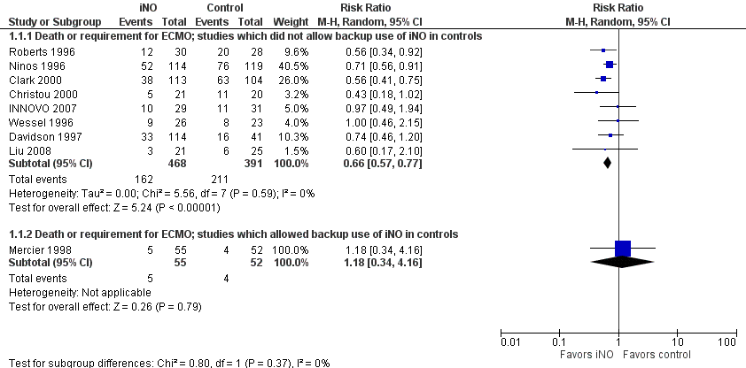
In general, to be confident about the impact of an intervention, it is preferable to see at least one very large trial with narrow confidence intervals including babies from multiple centers. The alternative being several trials with different risks in the controls and similar relative risk reductions in order to conclude that something which appears effective in a very high risk population is also effective in a lower risk population.  That is the kind of information that we have for inhaled nitric oxide in term infants, for example. Here is the Forest plot for the iNO studies, as you can see, the larger studies had relative risks of “death or ECMO” between 0.56 and 0.74, and the confidence intervals all overlap substantially, despite differing control group risks.

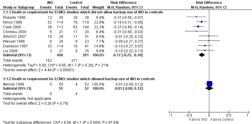
The second figure is revised from the the term iNO Cochrane review, I have ordered the studies by the control group risk of adverse outcome, and shown the risk difference. As you might expect, the higher the control group risk, the greater, in general, is the risk difference, even though the relative risk, (or risk ratio) is similar. That is why you need to be sceptical when you hear statements such as “it has only been shown to work in high-risk groups”, as we used to hear about probiotics. It is much harder to show a “statistically significant” outcome when the baseline risk is lower. But if the relative risk reductions are similar across studies with different baseline risk, that is a good reason to think that the impacts are the same.

For this positioning intervention, which has significant impacts on care of these babies, the high incidence of PIVH in the controls, reducing to an incidence with the intervention which is more similar to other recent publications, is consistent with an impact of the intervention, but is also consistent with a randomly higher incidence in the controls than usual. This is why to me the meaning of the ‘18% of these infants’ is important, if, over several years, they have had an 18% incidence of severe IVH among the ELBW, and in this study it is similar at 20%, with a substantially lower incidence in the group who had the intervention, I think that is more likely that the reduction is a real impact of the intervention, (in contrast to having a usual rate of 7.2% that increased to 20% during the period of the study).

So what do other trials say? Well there aren’t any, I think. The 2017 Cochrane review found 2 trials comparing supine positioning with the head midline, to supine with the head turned to the side (total n=110), and found no differences of note in any outcome.

A potentially important study, which certainly needs to be repeated, and it would be nice to study head elevation and midline positioning as separate interventions.

The other trial that has made me think about this issue is the PREMILOC trial. (Baud O, et al. Effect of early low-dose hydrocortisone on survival without bronchopulmonary dysplasia in extremely preterm infants (PREMILOC): a double-blind, placebo-controlled, multicentre, randomised trial. The Lancet. 2016;387(10030):1827-36). In that trial, routine administration of hydrocortisone at low doses over a total of 10 days led to a reduction in mortality before discharge, and a reduction in oxygen use at 36 weeks compared to control in infants of 24 to under 28 weeks.

When trying to assess the relevance for my practice, I note that the study did not include any infants under 24 weeks, or with a birth weight below the 3rd percentile, or with ruptured membranes before 22 weeks, or with 5 minute Apgar under 4, or with congential anomalies detected prenatally. It was overall a very high quality multicenter trial including a little over 1000 babies.

One of the striking things in the results of that trial is the high mortality before discharge in the 24 and 25 week infants, which is 44% with placebo and 42% with hydrocortisone. In my practice and in the 2017 CNN report, mortality among infants at 24 and 25 weeks admitted to the NICU (data which include those other very high risk groups) is 20%. The incidence of severe IVH is also very high in PREMILOC  at about 25%. Even at 26 and 27 weeks mortality in both groups is higher than the CNN mortality, 7.6% HC and 15.3% placebo in the PREMILOC trial, compared to 6% in the CNN, again including growth restricted babies.

There are also other signs in the publication that the treatment approaches were substantially different to what we do in our practice, 35% of placebo and 30% of hydrocortisone babies were on inotropes at study entry, 43% in each group received insulin during the trial.

If I already have a mortality which is substantially lower than the intervention group in a trial such as this, am I likely to see an impact of the intervention in my practice?

The answer, I would say, is definitely a ‘maybe’. If the same pathophysiology for the adverse outcome exists, and if the underlying risk factors are similar (including aspects of obstetric management), then I might expect a similar relative reduction in mortality, and a smaller absolute reduction in mortality. Of course as the absolute benefit become smaller, the potential secondary adverse effects of the intervention become potentially more important, in this specific case the increase in late-onset sepsis may be more important in the calculation of the risk-benefit balance.

I am not saying that we should ignore the results of trials if there are details of management or outcomes that are different to local data, that would just lead to chaos and a complete rejection of evidence-based practice, I am saying that we need to be thoughtful about interpretation and application of trial data. If local approaches and outcomes are dramatically different from a published trial, the we should take into account the toxicity/adverse impacts of the intervention, and whether there are other data from a variety of populations that are consistent. To go back to the probiotics example, the relative reduction in the risk of NEC is similar across studies, suggesting a real impact overall, although there are differences between the studies they consistently found no adverse effects of priobiotic use. Even if you have locally a low incidence of NEC, it is likely that it will be further reduced by probiotics, without any increased risk, and without much cost.

For routine midline head-elevated positioning, or routine use of hydrocortisone in the 1st day of life, I will wait for more data from different groups with differing baseline risks before changing my practice.

Posted in Neonatal Research | 1 Comment

Pulse Oximetry screening; a bizarre decision in the UK.

Universal pulse oximetry screening for critical congenital heart disease is a simple cheap addition to universal hearing and metabolic screening with undeniable benefits. Infants with undiagnosed life threatening congenital heart disease can be detected prior to closure of the ductus arteriosus, and prior to discharge from hospital. Infants who have such critical disease can have intervention, including surgery, with a lower mortality compared to infants who present after discharge who are often in shock at the time of diagnosis. Many bodies, including the Canadian Pediatric Society have come out in favour of universal pulse oximetry screening as a result.

Despite all of the data about the beneifts of screening, in the UK the National Screening Committee has just recommended against inclusion of pulse oximetry screening in the national program of neonatal screening, which basically means the neonatal physical exam.

The main justification of this decision appears to be their evaluation of evidence about “harms” of screening. Potential harms are listed in the following way:

• A positive result from pulse oximetry will generate some harms, including: parental anxiety, a longer stay in hospital, possible transfer to the neonatal unit, further tests to assess for non-symptomatic conditions.
• For many of these babies the further investigations will be unnecessary and the baby will be identified as healthy. This is a false positive result.

They also seem to doubt the benefits of a true-positive screen:

• For babies with CHD or other non-cardiac condition it is not clear that investigations and identification of these conditions will lead to any better outcome than a diagnosis at the time the baby becomes symptomatic.

I find this decision bizarre given that the preferred method of the committee is therefore physical examination, which has a false negative rate in the UK of over 50%, and a false positive rate of about 50%. Those data are referred to in this report, as the best studies were performed in the UK, but the potential harms of physical examination (false positive rate high) and false reassurance of negative physical examinations is not considered. I think the routine physical examination is therefore far more questionable than routine pulse oximetry, for the detection of congenital heart disease.

In some studies, in fact, referral for neonatal cardiac ultrasound is not increased by routine pulse oximetry screening. It is just more appropriately targeted. As about 4 to 5% of newborn infants will have a murmur, referral based on the clinical exam leads to many more false positives, especially when the target condition is critical heart disease, than oximetry.

What I find most bizarre, is that between the actual report of the literature review, and the chapter which compares the findings of that review to the criteria for institution of a screening program, there is a huge disconnect. The addition of pulse oximetry screening to the neonatal exam seems to fulfill all the criteria in the list.

Criterion 5

This risk of discharge home without a diagnosis or of severe acidosis has been estimated to be reduced by around 60% with pulse oximetry.
The benefit of newborn screening will be reduced if antenatal detection increases significantly, however current models suggest that newborn screening will remain clinically effective and cost-effective for life-threatening or critical CHDs until antenatal detection rates are above 85-90%…
Non-cardiac conditions leading to low oxygen saturation, such as respiratory or infective illness, may be found in infants with low oxygen saturations (false positive screening results). The benefits and costs of further investigation and early diagnosis of such conditions requires further investigation before these diagnoses can be considered a benefit of screening.

I do agree with that last paragraph, babies with lowish saturations who have, for example, increased pulmonary vascular resistance may do well without intervention, just being the slowest percentiles for the resolution of their fetal pulmonary vasoconstriction. Some probably do benefit from finding low saturations, such as those with sepsis, but it isn’t clear from the literature exactly how many “false positives” actually have conditions that need, and benefit from, intervention. But I also don’t think you can write all the ‘false positives’ off as an undue risk of screening, at least some of the babies will benefit.

The consultation process included an in-depth examination of the ‘false positives’ from the UK pilot study. you can see that if you click on the ‘Notes from the Pulse Oximetry workshop’ on this page. That review examined what happened to the 239 babies with a positive screen (0.73% of the screens performed); there were 14 babies with congential heart disease. Being fairly conservative in their opinions, there were another 36 babies who had conditions requiring treatment. 32 babies had discharge delayed despite not having a treatable condition, and the remaining false positives went home as planned anyway.

Criterion 8

A pathway for clinical investigation after a positive screen result on pulse oximetry has not been clearly established or evaluated in practice. Essential considerations prior to implementation of pulse oximetry in a national screening programme would therefore be to agree a policy for
investigation to identify cardiac and non-cardiac causes of low oxygen saturation, including consideration of the resource implications and acceptability to parents.

Not really in agreement here, the first stage of evaluation of a baby with a confirmed positive screen should be a rapid expert cardiac ultrasound, if there is no structural heart disease, then the second step is not entirely clear, I would agree. Who needs evaluation in what order for what conditions? Do they all need a chest x-ray? Or blood culture) But eliminating critical congential heart disease is a clear priority as the first step on the pathway.

Criterion 10

…Early detection of life-threatening CHDs in asymptomatic newborns allows management aimed at preventing cardiovascular collapse before intervention, a particular risk for duct-dependent cardiac defects, and there is some evidence that this can lead to improved short and long-term outcomes after surgery.

Yep.

Criterion 14

Antenatal ultrasound, newborn clinical examination and pulse oximetry appear acceptable as  screening tests. However the acceptability of high false positive rates (which may raise anxiety) and  false negative rates (leading to false reassurance) requires further exploration for all screening modalities.

I don’t know the literature about parental anxiety from false positive oximetry screening, but there are several studies about parental impacts of false positive hearing screens, which are very reassuring. They show that false positive screens are not a major burden to families, and that they appreciate the value of screening despite the false positive test of their child. It is important to have good communication with the parents prior to or during the screen, with written and/or verbal information. The experience from the Birmingham study seems to show the same thing. (Powell R, et al. Pulse oximetry screening for congenital heart defects in newborn infants: an evaluation of acceptability to mothers. Archives of disease in childhood Fetal and neonatal edition. 2013;98(1):F59-63).

Most neonatal screening tests are trying to detect relatively rare phenomena, and false postives for almost everything (which may cause stress) are more common than true positives. In this light, the ratio of true positives to false positives for pulse oximetry screening is similar to many of the things we already screen for. For example for neonatal hypothyroidism screening there are between 2 to 3 times as many false positives as true positives, and if your program starts with T4 screens that ratio is even higher. The benefits of hypothyroidism screening are so evident that we accept a substantial number of false positives, and so do the parents.

As for false negatives, this is also an issue with other screens, such as hearing screening, and parental information is key. But is it truly an issue? If a child has critical congenital heart disease which is missed by a falsely negative oximetry screen, and then presents with a serious deterioration later on, are they worse off than if they had not had a screen at all? Are parents likely to say “he had a normal oximetry screen, so we won’t take him to the emergency room?” I think kids with a false negative screen will likely be in exactly the same position as non-screened kids, therefore without an adverse impact.

Criterion 15

Existing evidence suggests that the benefits outweigh the harms for newborn screening, when the screening test is clinical examination with or without pulse oximetry, and for antenatal screening, when the screening test is antenatal ultrasound.

Agreed

Criterion 16

The existing evidence strongly suggests that pulse oximetry in conjunction with clinical examination is more cost-effective than clinical examination alone. Further evidence, including estimation of  QALYs, continues to support this.

Strongly agreed!

The literature review does have a couple of strange features, including a figure showing a list of congenital heart diseases causing death in the first year of life, which is headed by ventricular septal defect. They don’t give the correct reference for the data from which the data are derived, and I can’t find any data in the references by Wren, that they seem to be referring to, that supports that figure. If the quality of the understanding of the problem is reflected by that figure (when did you last see an infant with a VSD die? Before 1 year of age? After excluding complex cardiac malformations which happen to include a VSD?) A fairly recent study from France, for example put VSD as the least likely to cause death before 1 year of age.

Overall I think the recommendation of this committee doesn’t follow from their review of the literature and their own pilot project. If you are in the UK you can make a comment before this is finalized, go the university of Birmingham website here, and follow the links.


Posted in Neonatal Research | Tagged , | 1 Comment

Gastric acid is good for your bones.

We’ve known for a while now that suppressing gastric acid production in preterm infants increases Necrotising Enterocolitis and also systemic sepsis. Presumably this is because the intestinal microbiome is deranged by allowing the survival of pathogens as they pass through the stomach. There is direct evidence of this effect.

In older children we also know from a large multicenter RCT that PPI (proton pump inhibitor) use increases pulmonary infections in children with poorly controlled asthma, without any benefit.

There are also data about the adverse impacts of PPI use on iron and calcium absorption, mostly from adults, and that this increases the chance of having low bone density and more fractures. They also cause hypomagnesaemia and reduce B12 absorption.

Now observational data from a huge database (Malchodi L, et al. Early Acid Suppression Therapy Exposure and Fracture in Young Children. Pediatrics. 2019:e20182625)
show the same association in young children. There were nearly a million children in the database of the US Military HealthCare System, and, amazingly, more than 10% of them had acid suppression therapy in the first year of life. 0.9% of the children had a PPI, 8% a H2Receptor Antagonist, and 2% had both.

Exposed infants were more likely to have fractures,  the longer they received acid suppressants and the earlier they started, the greater the risk.

Although there is some doubt about the precise mechanism, the data are very consistent, and our very preterm babies, who are already at risk of poor bone mineralisation, should not be placed on acid suppression therapy unless there is reliable evidence that they have a clinical condition caused by gastric acid.

For most of the 10% of the infant population who received the therapy in this study, I can wager that there was no such evidence apart from, perhaps, a tiny minority. For nearly all of the NICU patients who receive such therapies, I can make the same wager, and be quite sure that my stake will be safe.

Even among infants who are thought to have clinical events related to reflux (a thought which is almost always shown to be erroneous when tested objectively) there is little or no reason to block acid production.

The latest in a large number of studies attempting to elucidate the relationship, if any, between reflux and cardio-respiratory events, presents recordings of multi-channel impedance, ph-metry and cardiorespiratory recordings in 52 preterm infants who had symptoms thought to be due to reflux (regurgitation, rumination or irritability) and on-going cardiorespiratory events. (Nobile S, et al. Correlation between cardiorespiratory events and gastro-esophageal reflux in preterm and term infants: Analysis of predisposing factors. Early Hum Dev. 2019;134:14-8.) Babies were on average about 1kg birth weight and between 1 and 2 months old when studied. Five of the 52 babies perhaps had a temporal association between reflux and events, as calculated by the “symptom association probability” of more than 95%, and in only 3 of those did the events follow the reflux, within 2 minutes. Of those events which followed reflux episodes, many were non-acid or very weakly acidic.

So even among the small proportion of events which occur within a couple of minutes of a proven reflux event, which may be a random association or possibly causative, there is no good reason to suppose that blocking gastric acid production will make any difference.

In the absence of multiple intraluminal impedance studies, the only clinical sign which strongly suggests reflux is a vomit stain somewhere. (As we are just past father’s day, I can testify to the diagnosis of reflux from the puke-stained shoulders of my shirts). If the baby is found with regurgitated milk in their bed, then they have had a reflux episode, no other clinical sign is reliable.

In a recent review article from an expert in the field, Sudarshan Jadcherla, the highlight of the conclusion regarding therapy of reflux disease was “No single pharmaceutical or non-pharmacological target exists to treat infant GERD”. Not only that but commonly used therapies are toxic, and may affect your patients’ bones.

Posted in Neonatal Research | Tagged , | 1 Comment

What respiratory outcomes are important?

When bronchopulmonary dysplasia was first described by Northway in 1967 he didn’t try to produce a definition, his paper was a description of a small number of preterm survivors of high oxygen and positive pressure ventilation. He noted some years later the long term implications for pulmonary health of the survivors, during adolescence and as young adults, many had hyperinflation, and small airways dysfunction was common. Many had on-going respiratory symptoms.

Since then we have been trying to arrive at a way of predicting long term pulmonary health without waiting for those very prolonged outcomes. Andy Shennan came up with the idea of oxygen requirements at 36 weeks because that was a better predictor of post-discharge pulmonary problems than oxygen need at 28 days, which had become the rather arbitrary definition prior to his publication. Even with the first publication in 1988, the positive predictive value of O2 at 36 weeks for post-discharge respiratory problems was only 63% and the NPV was 90%. With changes in survival and the pattern of lung injury, the usefulness of oxygen at 36 weeks as a predictor of clinically important pulmonary injury has diminished.

A study by the Canadian Neonatal Network/Neonatal Follow-up Network examined the predictive ability of various combinations of oxygen therapy and other respiratory support among infants <29 weeks gestation for predicting what they called “serious respiratory morbidity” which was defined as either (1) 3 or more rehospitalizations after NICU discharge owing to respiratory problems (infectious or noninfectious); (2) having a tracheostomy; (3) using respiratory monitoring or support devices at home such as an apnea monitor or pulse oximeter; and (4) being on home oxygen or continuous positive airway pressure at the time of assessment between 18 and 21 months corrected age. (Isayama T, et al. Revisiting the Definition of Bronchopulmonary Dysplasia: Effect of Changing Panoply of Respiratory Support for Preterm Neonates. JAMA Pediatr. 2017;171(3):271-9.) At least 3 rehospitalizations was chosen because the 95th percentile of the number of readmissions owing to respiratory problems in this cohort was 2. Such serious morbidity occurred in 6% of the approximately 1500 babies included, and was not well predicted by oxygen requirements at 36 weeks, it was better predicted by either oxygen or respiratory support (which included high flow nasal cannulae at more than 1.5 litres per minute or other invasive or non-invasive support) and determining that at 40 weeks post-menstrual age was a better predictor than at 36 weeks. 24% of the babies in the cohort had oxygen or respiratory support at 40 weeks, of them 16% had “serious respiratory morbidity”, of those who did not have O2 or respiratory support at 40 weeks 2.4% had this severe morbidity.

Although this definition gives an adjusted odds ratio for serious respiratory morbidity of 6.1 (95% CI 3.4-11.0), which is better than a definition at 36 weeks, I am not sure how useful it is, with a PPV of 16% and a fairly high negative predictive value, a large majority of infants with this definition of BPD do not have ‘serious respiratory morbidity’.

The bigger problem maybe, is the definition of SRM (typing the whole phrase takes me too long!) being home on an apnea monitor doesn’t seem equivalent to me to having 3 hospitalisations, or still being on CPAP at 18 month follow-up. I think determining which definition of BPD, (a definition created by physicians), is best at predicting SRM, as defined by physicians, won’t necessarily help us a great deal. We should first determine which respiratory outcomes are most important to families, and then rank the clinically important outcomes (or rather ask parents to rank them), if we did that we could produce some sort of ordinal score of severity of preterm chronic lung disease. Then we could figure out if it is possible to predict those outcomes before they occur. We also should be very careful about introducing items which may be very strongly affected by different practice patterns, (such as home apnea or saturation monitoring), in some centers home use of such monitors is exceedingly rare, compared to others.

If I were to guess, I would think that families, during the first year or 2 or life, would be more disturbed by emergency room visits, and hospitalisations (especially PICU re-hospitalisations) and next by home oxygen, especially as the infant becomes more active, and next by daily use of respiratory medications. Tracheostomy would probably be way out at the top, but includes very few babies in Canada. But I don’t think my guesses are worth much, we should really ask parents, and other members of society.

Another recent publication from the NICHD network only examined possible variations of BPD definitions at 36 weeks, not at other post-menstrual ages. (Jensen EA, et al. The Diagnosis of Bronchopulmonary Dysplasia in Very Preterm Infants: An Evidence-Based Approach. Am J Respir Crit Care Med. 2019)  They tested 18 different ways of analyzing different levels of respiratory support and FiO2 at 36 weeks for their accuracy in predicting post-discharge serious respiratory morbidity which was defined as death between 36 weeks and follow up or:

the occurrence of at least one of the following: tracheostomy placed any time prior to follow-up; continued hospitalization for respiratory reasons at or beyond 50 weeks PMA; use of supplemental oxygen, respiratory support, or respiratory monitoring (e.g. pulse oximeter, apnea monitor) at follow-up; or ≥2 re-hospitalizations for respiratory reasons prior to follow-up. Continued hospitalization at 50 weeks PMA is approximately 2 standard deviations above the mean age at discharge for extremely preterm infants included in Neonatal Research Network studies. Two or more re-hospitalizations represents the upper 75th percentile for re-hospitalization number among Network babies.

In this study, the best definition appeared to depend solely on the level of respiratory support at 36 weeks irrespective of the oxygen needs, using these diagnostic criteria, infants breathing in room air at 36 weeks PMA did not have BPD. Disease severity among the remaining infants was classified according to support: grade 1, nasal cannula at flow rates ≤2L/min; grade 2, nasal cannula at flow rates >2L/min or non-invasive positive airway pressure; and grade 3, invasive mechanical ventilation.

Using these definitions the percentage of babies with respiratory morbidity increased from 10% among infants without BPD, to 19% with grade 1, 35% with grade 2, and 77% with grade 3.

My comments about this study echo those for the CNN paper, the relative importance of these different aspects of SRM is variable, and an overall moderately good capacity to predict which infants may develop SRM is interesting, but would it be enough to be used as an interim outcome measure for clinical trials? Or as a marker of respiratory outcomes for quality control? In addition some of the measures are likely to be heavily influenced by practice patterns, some centers will use higher flow rates and lower oxygen concentrations, some will remove CPAP earlier than others, or extubate with higher ventilatory requirements.

One of the reasons for bringing this up now is a publication of a study aimed at reducing lung injury in the very preterm infant. Davis JM, et al. The role of recombinant human CC10 in the prevention of chronic pulmonary insufficiency of prematurity. Pediatr Res. 2019. I think this is the second small preliminary trial of Clara Cell Protein in very preterm babies, the previous one having 22 babies total (7 controls, 7 with low dose and 7 with higher dose Clara Cell Protein, called CC10 in the new study). It is a recombinant version of  a protein produced by human Clara cells, which has several potentially beneficial effects, including reduction of lung inflammation. In the previous trial there was a reduction of signs of lung inflammation with a single intratracheal dose, that study was underpowered for clinical outcomes, and didn’t really show any evidence of clinical benefit. The new trial included 44 babies in all, of 24 to 29 weeks and intubated for RDS. They were included in 1 of 2 sub-studies, the first randomized kids to low dose (1.25 mg/kg) or placebo, the second to higher dose (5 mg/kg) or placebo, given as a single intratracheal dose within 4 hours of surfactant, and at less than 24 hours of age.

The primary outcome of the study was survival without what they called CPIP, chronic pulmonary insufficiency of prematurity, which was defined as presence of any one of the following : (1) evidence of respiratory symptoms (e.g., coughing and wheezing) or use of respiratory medications by parental diaries or pulmonary questionnaires (CPIP-SS), (2) one or more re-hospitalizations for respiratory causes (CPIP-RH), (3) administration of respiratory medications (including oxygen) (CPIP-RM), and (4) at least one non-routine medical visit for respiratory causes (CPIP-DV).

These sound like things that are likely to be of some importance to families; the primary outcome was analyzed as a dichotomous, yes/no outcome, and in addition they recorded the number of criteria for CPIP that were satisfied.

These criteria are clearly much less severe than the outcomes of the 2 other studies discussed above, and almost all of the babies had CPIP, with no difference between placebo and CC10 groups, at either dose. Overall only 8 of the 44 babies survived without CPIP. About half of the babies in the trial did not have BPD by the usual criterion of oxygen needs at 36 weeks, and most of the babies who did not have BPD still had CPIP.

What should the outcome of interest be for studies trying to reduce lung injury in the very preterm infant? Is an interim outcome such as BPD, by any of these definitions, of sufficient predictive capacity to use it in place of clinically important pulmonary dysfunction in the first years of life? I think we have to be very careful that, even if we could come up with criteria that predict chronic pulmonary symptoms among the general preterm population, an intervention that acutely reduces the chances of satisfying those criteria will not necessarily improve long term pulmonary function. Post-natal steroids, for example, by reducing inflammation, reduce the number of babies who satisfy definitions of BPD which are based on oxygen needs, but there is no evidence that they improve long term pulmonary function.

In order to answer the question of the usefulness of “BPD” as a surrogate outcome for improvements in pulmonary health in clinical trials, Anna Maria Hibbs and her colleagues have performed a systematic review of large trials of BPD prevention that also published data on long term respiratory health outcomes Corwin BK, et al. Bronchopulmonary dysplasia appropriateness as a surrogate marker for long-term pulmonary outcomes: A Systematic review. J Neonatal Perinatal Med. 2018;11(2):121-30. The review found 5 trials, the DINO trial from Melbourne of DHA for preventing developmental impairment, which also looked at lung injury, the NO-CLD trial, the early iNO trial which disappointingly has no obvious acronym, SUPPORT, and the SOD the lungs trial (that is actually my own personal acronym for the trial).

All the studies reported BPD, using a variety of definitions, and all had some measures of longer term pulmonary health: all reported healthcare utilization,  (hospitalizations, and visits to the doctor or the emergency room); 3 reported respiratory illness, (e.g. asthma); 4 reported respiratory medication use (e.g. bronchodilators, steroids, diuretics, oxygen); one study reported 1 year mortality.

A consistent relationship between BPD and subsequent markers of respiratory morbidity was not seen in the trials studied. Only the NO CLD trial found a significant decrease in rates of BPD. This study also saw significant decreases in the use of respiratory medications including oxygen, but in no other outcome measures. The SOD study also found a significant decrease in respiratory illness requiring asthma medications, but failed to find a significant decrease in BPD. Similarly, the SUPPORT study failed to find a significant decrease in BPD, but did see significant decreases in measures of healthcare utilization and respiratory illness.

Not only therefore is BPD, using any definition, a very limited way of describing severity of early pulmonary injury, it is clearly of very low value as a surrogate for clinical trials aimed at improving pulmonary health.

This brings into even sharper relief the concerns about using BPD as part of the combined outcome of BPD or death. Mortality matters, BPD, not so much.

Chronic pulmonary dysfunction and respiratory illness in early life are important adverse outcomes for families, finding better ways to describe, predict, and prevent them is a priority.

Posted in Neonatal Research | Tagged , , , | 1 Comment

High-flow in non-tertiary neonatal units: Hunting for answers. #EBNEO

I think Brett Manley is going for the record as the person with the highest proportion of his publications in the FPNEJM, he now has 3, with 2 of them as first author. This is the HUNTER trial where babies in level 2b NICUs, as we could call them, i.e. nurseries with access to CPAP, but not prolonged invasive ventilation, were randomized to receive either CPAP or high-flow nasal therapy. (Manley BJ, et al. Nasal High-Flow Therapy for Newborn Infants in Special Care Nurseries. The New England journal of medicine. 2019;380(21):2031-40) Babies are not usually kept in such units if they are less than 32 weeks or less than 1200 g, so to be eligible for this trial they had to be at least 31 weeks gestation, >1200 g birth weight, and to need non-invasive respiratory support according to the attending pediatrician. This was performed as a non-inferiority trial, and was designed to be able to detect an increase in therapeutic failure from 17% with CPAP to 27% with high flow.

Babies in the high flow group were placed on 6 litres per minute of a heated humidified gas mixture via the Fisher-Paykell Optiflow device, which could be increased to a maximum of 8 lpm. CPAP was delivered using a bubble system and either a mask or prongs, at 6 cmH2O, which could be increased to a max of 8 cmH2O. If a high-flow baby failed they could be treated with CPAP at 8 cmH2O, if a CPAP baby failed they were out of the trial and a discussion with the regional NICU was expected.

There were over 750 babies randomized, and the primary outcome was treatment failure in the first 72 hours: which was defined as; if they got to maximal support and needed more than 40% oxygen for more than 1 hour (to stay at 91 to 96% saturation), or had a respiratory acidosis to less than 7.2, or had severe apnea.

Treatment failure occurred in 20.5% of the high flow babies and 10.2% of the CPAP group. Many of those who failed high flow were transferred to CPAP and about half of them stabilized, so in the end just over 5% of each group were intubated within the 1st 72 h, and around 6% in total in each group; there were twice as many pneumothoraces needing intervention in the CPAP group (4.8% vs 2.4%, 95% confidence intervals include compatibility with no difference). Slightly more babies in the high-flow group were transferred to tertiary NICUs, 13% vs 11%.

This is really important clinically relevant data for level 2 nurseries. The eventual clinical outcomes were quite similar in the two groups, so I think it would be acceptable to continue to use high-flow in such nurseries as long as you had back up CPAP readily available. There were  no clear advantages of high-flow demonstrated in this study but other studies have found that parents prefer high-flow, and they appear to be more comfortable for the babies.

The only quarrel I have with this study is how tortuous the language becomes with non-inferiority studies. We are told that high-flow was “not non-inferior” to CPAP. Why not just “was inferior”? There were substantially more treatment failures, which passed the non-inferiority margin, hence the intervention was inferior in terms of the primary outcome.

Posted in Neonatal Research | Tagged , , , | 1 Comment