The preterm GI tract is not sterile

The FDA are at it again, they seem to be on a mission to go after suppliers of probiotics for preterm babies, and have now attacked Abbott. They appear to have demanded that they stop marketing their probiotic product, as it is not an approved medication, and could be considered an adulterated food product that is not GRAS (Generally Regarded As Safe).

I don’t know if it will be possible to get the FDA to back off. They need to realize that all breast-fed babies are already receiving probiotics. In a completely unregulated way. Every baby that gets unpasteurized breast milk is getting some sort of bifidobacteria, and probably lactobacilli, as well.

If breast-milk isn’t GRAS, I don’t know what is!

Bifidobacteria are usually present in fresh breast milk, although sometimes in very small amounts, and mixed with a huge variety of other organisms, the microbiome of milk varies between individuals and varies around the world, and it seems to be changing over time. Even among babies whose mothers’ milk contains few bifidobacteria, the infants become overwhelmingly colonized with bifidobacteria within a few days, for as long as they are breast fed. Babies born by cesarean delivery, and/or who receive antibiotics in the neonatal period, develop a very different microbiome, which I don’t think is too far of a stretch to call “abnormal”; there is an increased frequency and relative abundance of various pathogens, including E. Coli, Klebsiella, and others.

Once a baby is admitted to the NICU, if we consider the preterm at risk of NEC, intestinal bacterial colonization proceeds with the organisms present in the environment, on the equipment, and in their feeds.

I think of administering probiotics, Florababy(TM) in the case of our NICU, as an attempt to push the microbiome towards normality. I know that despite routine administration of probiotics, the microbiome of the babies in my NICU will remain abnormal, and we will still have cases of late-onset sepsis and NEC. Even, occasionally, of sepsis caused by the organisms that we give purposefully. But we cannot avoid giving the babies enteral organisms! They will become colonized whatever we do, and without probiotics the balance will be more towards pathogens.

Nolan LS, et al. The Role of Human Milk Oligosaccharides and Probiotics on the Neonatal Microbiome and Risk of Necrotizing Enterocolitis: A Narrative Review. Nutrients. 2020;12(10).

The best way to nudge the intestinal microbiome towards being normal is to :

  1. Ensure that all the babies receive unpasteurized mother’s own milk as soon as possible after delivery. There may be additional benefits of using colostrum for the first feeds.
  2. Avoid antibiotics, or limit them to the fewest babies for the shortest time possible
  3. Continue to feed with mother’s own milk, or if unavailable/insufficient, use donor human milk
  4. Administer a high-quality probiotic preparation. My best guess is that it should contain B longum ssp infantis, and at least one other organism, perhaps Lactobacillus rhamnosus

Adding human milk oligosaccharides, HMOs, especially DSLNT (disiallylo-N-tetraose) improves colonization with Bifidobacteria, and further normalises the microbiome. It is fascinating to reflect on the fact that human milk contains oligosaccharides that humans cannot metabolise! They make up a major proportion of the solids in breast milk. Bifidobacteria have a unique pathway, the Fructose-6-Phosphate Phosphoketolase system, that allows them to metabolize those HMOs, and as a result to downregulate inflammation. They create communities in our guts where multiple species co-operate, which has been referred to as “altruistic” behaviour.

In the future, I think that additional specific HMOs will probably be added to my list of microbiome interventions; if the FDA permit it.

One thing we cannot do, and should not try, is to keep the preterm infant’s GI tract sterile. Trying to ensure the most normal possible microbiome is an essential part of care of the extreme preterm. The FDA’s interventions will only ensure that intestinal colonization is more random, with more pathogens, and more cases of NEC will follow. The FDA seem really to want to kill preterm babies. The lack of insight into the impact of this intervention is startling.

As far as I can see, there is no current pathway for the approval of probiotics for administration to preterm infants. It should be a major priority of the FDA to create and facilitate such a pathway, this is an urgent need for preterm babies. And to back off from those who are currently supplying high-quality products in the interim.

Here are some of the references I used for this post.

Nolan LS, et al. The Role of Human Milk Oligosaccharides and Probiotics on the Neonatal Microbiome and Risk of Necrotizing Enterocolitis: A Narrative Review. Nutrients. 2020;12(10).
Egan M, Van Sinderen D. Carbohydrate Metabolism in Bifidobacteria. The Bifidobacteria and Related Organisms. 2018. p. 145-64.
Moossavi S, et al. Composition and Variation of the Human Milk Microbiota Are Influenced by Maternal and Early-Life Factors. Cell Host Microbe. 2019;25(2):324-35 e4.
Henrick BM, et al. Elevated Fecal pH Indicates a Profound Change in the Breastfed Infant Gut Microbiome Due to Reduction of Bifidobacterium over the Past Century. mSphere. 2018;3(2):10.1128/msphere.00041-18.
Kumar H, et al. Distinct Patterns in Human Milk Microbiota and Fatty Acid Profiles Across Specific Geographic Locations. Front Microbiol. 2016;7:1619.
Jeurink PV, et al. Human milk: a source of more life than we imagine. Beneficial microbes. 2013;4(1):17-30.
Biagi E, et al. The Bacterial Ecosystem of Mother’s Milk and Infant’s Mouth and Gut. Front Microbiol. 2017;8:1214.
Notarbartolo V, et al. Composition of Human Breast Milk Microbiota and Its Role in Children’s Health. Pediatr Gastroenterol Hepatol Nutr. 2022;25(3):194-210.
Chang CM, et al. Effects of Probiotics on Gut Microbiomes of Extremely Preterm Infants in the Neonatal Intensive Care Unit: A Prospective Cohort Study. Nutrients. 2022;14(15).
Baucells BJ, et al. Effectiveness of a probiotic combination on the neurodevelopment of the very premature infant. Sci Rep. 2023;13(1):10344.
van Best N, et al. Influence of probiotic supplementation on the developing microbiota in human preterm neonates. Gut Microbes. 2020;12(1):1-16.
Larke JA, et al. Preterm Infant Fecal Microbiota and Metabolite Profiles Are Modulated in a Probiotic Specific Manner. J Pediatr Gastroenterol Nutr. 2022;75(4):535-42.
Saturio S, et al. Role of Bifidobacteria on Infant Health. Microorganisms. 2021;9(12).
Patangia DV, et al. Impact of antibiotics on the human microbiome and consequences for host health. Microbiologyopen. 2022;11(1):e1260.
Murphy K, et al. The Composition of Human Milk and Infant Faecal Microbiota Over the First Three Months of Life: A Pilot Study. Sci Rep. 2017;7:40597.

Posted in Neonatal Research | Tagged , , , , , | 4 Comments

How to express negative results… or positive ones

After any trial result, there is always a possibility that the true effect of an intervention is different to that shown in the sample who were studied. That is the whole rationale behind using statistics, a trial on a small sample will usually be compatible with a wide range of possible effects if the entire population had been treated. Exactly how to express the results of a negative trial is an ongoing debate.

Three simultaneously published trials in JAMA were all negative, that is they all showed no clear benefit of the intervention. The first was a multi-centre randomised trial in adults who are receiving assisted ventilation after a trauma (Albert RK, et al. Sigh Ventilation in Patients With Trauma: The SiVent Randomized Clinical Trial. JAMA. 2023). The intervention group had added sigh breaths, to reduce atelectasis, up to 35 cmH2O every 6 minutes. The primary outcome was ventilator-free days up to 28 days after admission, Which was scored as 0 if the patient died, and up to 28 if they were extubated immediately. The main results are presented thus

“The unadjusted mean difference in ventilator-free days between groups was 1.9 days (95% CI, 0.1 to 3.6) and the prespecified adjusted mean difference was 1.4 days (95% CI, −0.2 to 3.0). For the prespecified secondary outcome, patients randomized to sighs had 28-day mortality of 11.6% (30/259) vs 17.6% (46/261) in those receiving usual care (P = .05)”

The interpretation is that there might indeed be a benefit of sighs, based largely on the 28 day mortality outcome.

“…the addition of sigh breaths did not significantly increase ventilator-free days. Prespecified secondary outcome data suggest that sighs are well-tolerated and may improve clinical outcomes.”

The second trial was in adults with septic shock who were tachycardic (Whitehouse T, et al. Landiolol and Organ Failure in Patients With Septic Shock: The STRESS-L Randomized Clinical Trial. JAMA. 2023); there are some observational data to suggest that such patients benefit from slowing down the catecholamine induced tachycardia with beta-blockade. So they performed this multi-centre RCT of landiolol with the primary outcome of “the mean Sequential Organ Failure Assessment (SOFA) score from randomization through 14 days. Secondary outcomes included mortality at days 28 and 90 and the number of adverse events in each group.” There was no difference in the SOFA scores, but the trial was stopped as the mortality was somewhat increased with the beta-blocker

“The mean (SD) SOFA score in the landiolol group was 8.8 (3.9) compared with 8.1 (3.2) in the standard care group (mean difference 0.75 [95% CI, −0.49 to 2.0]; P = .24). Mortality at day 28 after randomization in the landiolol group was 37.1% (23 of 62) and 25.4% (16 of 63) in the standard care group (absolute difference, 11.7% [95% CI, −4.4% to 27.8%]; P = .16). Mortality at day 90 after randomization was 43.5% (27 of 62) in the landiolol group and 28.6% (18 of 63) in the standard care group (absolute difference, 15% [95% CI, −1.7% to 31.6%]; P = .08)”

Quite a large increase in mortality, in the “wrong” direction, but no “statistically significant” difference. Their interpretation:

landiolol “did not reduce organ failure measured by the SOFA score over 14 days from randomization. These results do not support the use of landiolol for managing tachycardia among patients treated with norepinephrine for established septic shock”

The third report is from the addition of 2 similar RCTs, in patients hospitalised with COVID, of the administration of vitamin C (Lovit-Covid Investigators, et al. Intravenous Vitamin C for Patients Hospitalized With COVID-19: Two Harmonized Randomized Clinical Trials. JAMA. 2023). Although previous investigations of Vitamin C use for critically ill patients have shown no benefit and its use has been largely abandoned, there was a SR and meta-analysis with a large number of tiny trials that showed the possibility of reduced mortality for COVID-19. Hence these two trials of intravenous vitamin C, one by the amazing Canadian Critical Care Trials group, the LO-VIT-COVID trial, and the other was the vitamin C arm of the REMAP-CAP trial “Both trials prospectively adopted the same intervention, outcomes, statistical analysis plan, and reporting, but the control groups were different. The LOVIT-COVID trial used a placebo for the control group and the REMAP-CAP trial used no vitamin C for the control group.”

The primary outcome was a composite of organ support–free days defined as days alive and free of respiratory and cardiovascular organ support in the intensive care unit up to day 21 and survival to hospital discharge. Values ranged from –1 organ support–free days for patients experiencing in-hospital death to 22 organ support–free days for those who survived without needing organ support.

I will reproduce the majority of the results section of the abstract here, I think it is a model of clarity.

Enrollment was terminated after statistical triggers for harm and futility were met.

Among critically ill patients, the median number of organ support–free days was 7 (IQR, −1 to 17 days) for the vitamin C group vs 10 (IQR, −1 to 17 days) for the control group (adjusted proportional OR, 0.88 [95% credible interval {CrI}, 0.73 to 1.06]) and the posterior probabilities were 8.6% (efficacy), 91.4% (harm), and 99.9% (futility). Among patients who were not critically ill, the median number of organ support–free days was 22 (IQR, 18 to 22 days) for the vitamin C group vs 22 (IQR, 21 to 22 days) for the control group (adjusted proportional OR, 0.80 [95% CrI, 0.60 to 1.01]) and the posterior probabilities were 2.9% (efficacy), 97.1% (harm), and greater than 99.9% (futility). Among critically ill patients, survival to hospital discharge was 61.9% (642/1037) for the vitamin C group vs 64.6% (343/531) for the control group (adjusted OR, 0.92 [95% CrI, 0.73 to 1.17]) and the posterior probability was 24.0% for efficacy. Among patients who were not critically ill, survival to hospital discharge was 85.1% (388/456) for the vitamin C group vs 86.6% (490/566) for the control group (adjusted OR, 0.86 [95% CrI, 0.61 to 1.17]) and the posterior probability was 17.8% for efficacy.

To clarify, the word “futility” has a definition in the statistical analysis section of the supplemental data, and has to do with the posterior probability of an advantage of vitamin C with an OR of >1.2 or more (I think), which these trials show is extremely unlikely. The first sentence of the discussion says it well:

In this large, harmonized, multinational randomized clinical trial, vitamin C administered to hospitalized patients with COVID-19 did not improve organ support–free days or hospital survival. On the contrary, there were high posterior probabilities (>90% for organ support–free days and >75% for hospital survival) that vitamin C worsened both outcomes in critically ill patients and those not critically ill.

As you can tell from the way the results are presented, these are Bayesian analyses, which give the probability of the real impact of an intervention, based on the prior probability (in this case, this was considered neutral) and the findings of the trial. Although there is overlap in the results from the 2 groups using traditional analysis, (“not statistically significant”), the Bayesian probabilities show it is unlikely that vitamin C is helpful, and most likely that it is, in fact, harmful.

The 3 trials are therefore reported as “no difference, but might be better than control”, “no difference, but might be worse than control”, and “probably worse, but almost certainly not better than control”. I must say I think that the Bayesian outcome presentation gives a better understanding of the likelihood that outcomes are worse with IV vitamin C. The other trials would have benefited from a posterior calculation of how likely it is that sighs improve survival (looks to be moderately likely, with a low likelihood of harm, I would guess), or how likely it is that beta-blockade is harmful (looks quite likely, and really unlikely to be beneficial). Also interesting is the primary outcomes used for the first trial. Duration of ventilator dependence and death are both part of the outcome, I am unsure how likely eventual survival is in adults who still need ventilation at 28 days after trauma, but you can see from these survival curves that there is almost no-one left intubated and alive by 24 days. This looks to me like a composite outcome that I could buy into, for this population.

Despite them being negative, or null trials, I think they will inform future practice, with sighs probably having a place in routine care of ventilated trauma patients, but not vitamin C for COVID, and especially not beta-blockade for tachycardia in septic shock.

The trial of late hypothermia among infants with HIE who didn’t arrive in time to start prior to 6 hours was also presented with a Bayesian analysis, which showed that, even though there was a null result by regular statistics, hypothermia was likely to be preferable for death or disability, with a posterior probablity of 76% of benefit. Laptook AR, et al. Effect of Therapeutic Hypothermia Initiated After 6 Hours of Age on Death or Disability Among Newborns With Hypoxic-Ischemic Encephalopathy: A Randomized Clinical Trial. JAMA. 2017;318(16):1550-60. That sort of analysis can gives us some confidence (an exact degree of confidence) that cooling is beneficial even if started a little after 6 hours.

Posted in Neonatal Research | Tagged , | Leave a comment

On which Planet? Reducing platelet transfusions in the newborn.

Recent publications about platelet transfusions in the newborn.

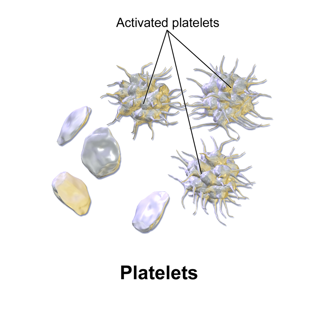
Hopefully, everyone has integrated the findings of Planet-2 into their protocols and guidelines. Curley A, et al. Randomized Trial of Platelet-Transfusion Thresholds in Neonates. N Engl J Med. 2019;380(3):242-51. This trial, in preterm newborns <34 weeks with any cause of thrombocytopaenia, apart from those with a recent major bleed, showed that a transfusion threshold of 25,000 led to fewer complications, including less bleeding, than transfusion at a more liberal threshold, of 50,000. The trial excluded babies who had a pre-existing “major IVH” which was defined by an intraventricular bleed with dilatation, or an intraparenchymal bleed of more than 2 cm maximum diameter on an ultrasound taken within 6 hours of randomization.

Picture copied from Wikipedia

A publication from our NICU (Zabeida A, et al. Platelet transfusion practice pattern before and after implementation of a local restrictive transfusion protocol in a neonatal intensive care unit. Transfusion. 2023;63(1):134-42) showed that our new guidelines, which mimicked the entry criteria for Planet-2, decreased the proportion of babies getting platelet transfusions, among all NICU admissions, from 9% to 5%, and eliminated the small number of babies who had previously sometimes received more than 3 transfusions. We were able to abide by the protocol for 70% of the transfusions, with some of the 20 transfused babies receiving transfusions at higher threshold because of active NEC, or being acutely postop. Three babies received transfusions because of low platelet counts (above the threshold) after ibuprofen treatment, and the concern that the ibuprofen causes platelet dysfunction, and there were 3 other protocol “violations” for different reasons.

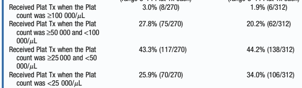
Another publication, this time from Bob Christensen’s group, (Bahr TM, et al. Platelet Transfusions in a Multi-Neonatal Intensive Care Unit Health Care Organization Before and After Publication of the PlaNeT-2 Clinical Trial. J Pediatr. 2023;257:113388) did not show a reduction in the proportion of babies receiving a platelet transfusion; 2/3 of the transfusions in their study, even after the guideline change, were given with platelet counts above 25,000. They did have some minor trends to reduced utilisation of platelets, (13 babies transfused per 1000 admissions compared to 16 before) but not what they were expecting.

These are the guidelines before and after the change

As you can see in this extract from their results table, most transfusions were still being given above 25,000

Another study, from a couple of years ago, also studied platelet transfusion before and after a more restrictive guideline, that guideline also has a threshold of 50,000 for infants considered to be at risk of IVH, (<28 weeks and <7 days). In contrast to the above study, they showed a dramatic reduction in platelet transfusions, from 25 per 100 admissions to 12. (Davenport PE, et al. Implementation of a neonatal platelet transfusion guideline to reduce non-indicated transfusions using a quality improvement framework. J Perinatol. 2021;41(6):1487-94), few of which were outside of their new guidelines. The justification given for the higher threshold in their guideline among infants at risk of IVH was “since the average time of randomization in the trial was day of life 7, and 39% of infants received a platelet transfusion prior to randomization, it was possible that these transfusions were given during the highest risk period for ICH”.

Long term follow up of the babies in the Planet-2 trial (Moore CM, et al. Two-year outcomes following a randomised platelet transfusion trial in preterm infants. Arch Dis Child Fetal Neonatal Ed. 2023;108(5):452-7) has now been published. The original publication reported death up to 28 days, but as far as I can see, nowhere does it report the total number of survivors per group, which should be mandatory, surely, for any RCT. The best I can find is a table in the supplemental materials, that shows the number of deaths which followed a Serious Adverse Event was 52 in the restricted, and 58 in the liberal transfusion group. In the 2 year follow up, the mortality before 2 years was 70 vs 91 (<25,000 threshold group vs <50,000 group), so there were either a lot of deaths after discharge, or a lot of deaths that were not considered a serious adverse event!!!

Among the survivors, there were a few more adverse outcomes for each outcome in the higher transfusion threshold group. There were 13% with CP compared to 10%, and slightly more with blindness, deafness, and seizure disorder. An important outcome which they reported was called “global developmental delay”, this was assessed variously, sometimes by formal testing, but often by an informal assessment conducted by a health care professional, who found the infant to be more than 9 months delayed in their development. I am very unsure how reliable this is as a method for determining “global developmental delay”, but I don’t think there is a good reason to suppose that the reliability would differ between groups; 44% vs 33% were given this appellation, with, again, the liberal, higher threshold, transfusion group being worse off.

There were also many more infants on oxygen (or respiratory support) at 2 years of age from the liberal transfusion group, 11% vs 4%. This is pretty serious lung injury, with more than twice as many infants being affected in the higher threshold group.

Given all of these adverse outcomes in the higher threshold transfusion group, and the lack of acute clinical benefit, you really need a good justification for transfusing above 25,000. It often takes a while to change clinical practice, and there may be occasional reasonable indications for transfusing earlier. Evidence-based protocols almost always improve practice and outcomes, they should be applied thoughtfully, and the impacts evaluated in day-to-day practice.

Given the lack of any benefit, and the higher risk of bleeding, with earlier platelet transfusions, and the concern that the results may not be applicable to babies at highest risk of IVH, or at-risk of extension of a previous major IVH, we need more trials. A trial that enrolled babies at birth, and/or which included babies with pre-existing severe intracranial haemorrhage, and/or examined even lower thresholds, would be feasible, and I think acceptable to many. One day we will know when to transfuse platelets, on this planet!

Posted in Neonatal Research | Tagged , , | 2 Comments

Addressing Prognostic Uncertainty

I recently gave a presentation to the Pediatrix consortium which was based on our review article about the approach to take after diagnosis of a serious intracranial haemorrhage (ICH) in the very preterm infant. (Chevallier M, et al. Decision-making for extremely preterm infants with severe hemorrhages on head ultrasound: Science, values, and communication skills. Semin Fetal Neonatal Med. 2023;28(3):101444)

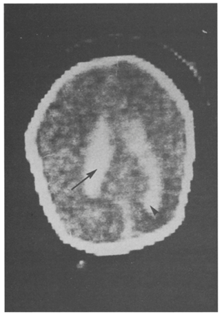
I first reviewed of the prognostic accuracy of severe ICH, making the point that there are 3 different types of haemorrhage which are all called grade 3. That is, those intraventricular haemorrhages that fill more than 50% of a lateral ventricle, those which acutely distend the ventricle, and those where there is blood in the ventricle and early post-haemorrhagic distension of the ventricle. These 3 types of grade 3 ICH may have different pathophysiology, and different prognostic implications, and, usually, it is not clear from a publication describing outcomes what they are including in their definition. The wording of the original system of classification by Lu-Ann Papile et al (based on CT scans) is somewhat ambiguous (“intraventricular hemorrhage with ventricular dilatation”), but the image was shown as an example in that publication, which I reproduce here, is of an ICH with acute haemorrhagic distension of the ventricle. In addition, bilateral and unilateral haemorrhages are put in the same category, and massive distension of the ventricles is lumped with those which have a minor degree of dilatation.

Grade 4 ICH, or intraparenchymal haemorrhage (IPH), is even more variable, with a small echodense spot being put in the same category, in almost all follow up studies, as massive bilateral bleeding with midline shift. There are systems to grade severity among IPH, (many of which are associated with ipsilateral lower grade haemorrhages, and may in that case be referred to as PVHI (peri-ventricular haemorrhagic infarction)). The Bassan system has been used the most. In that system, an ultrasound showing IPH is scored 0 or 1 for each of 3 features, IPH affecting more than one brain region, midline shift, and bilateral bleeding. Thus a grade 4 can have a score of 0, or up to 3.

This figure, form the Bassan study, shows how the brain regions are defined, based on imaginary lines drawn around the thalamus, and the relative proportions according to location.

There is never certainty in prognosis. That is a simplistic truism in all of medicine (or all of life!) but it is particularly true after ICH in preterm babies. Given the limited data we have, grade 3 hemorrhages, which are not accompanied by an IPH and are not followed by post haemorrhagic ventricular dilatation (PHVD), probably have little impact on development or motor function.

Grade 3 haemorrhages with subsequent progressive dilatation have impacts on motor function, and probably on language and cognitive development, but, in the absence of IPH these effects are relatively small, especially if early derivation is performed.

IPH outcomes vary between no impact and major global delay with tetraplegic CP. For an individual baby, the correlation between location and extent of the IPH on ultrasound and motor or developmental outcome is limited, and very variable in the reports. For example it has been reported that anterior IPHs are more likely to be associated with CP, or posterior IPHs, or that there is no effect of location. There seems to be an association between extent of the lesion and the presence of more severe developmental problems in the long term, and the Bassan classification does show some gradation in outcomes, IPH which score 0 or 1 having no major impact on long term outcomes, and those of 2 or 3 being associated with an increased risk of… you got it , “NDI”! In all the studies however, even the worst IPH may be followed by only mild long term abnormalities.

Several studies show that outcomes are affected more by other complications of neonatal care than they are by ICH. For example, this figure (Merhar SL, et al. Grade and laterality of intraventricular haemorrhage to predict 18-22 month neurodevelopmental outcomes in extremely low birthweight infants. Acta Paediatr. 2012;101(4):414-8) shows that a bilateral IPH in a baby who does not receive dexamethasone, and does not have an episode of sepsis, has a better prognosis (in terms of proportion with “NDI”) than a baby with bilateral sub-ependymal haemorrhage who has both of those factors.

What to do with these uncertainties? It would be simple to never make a decision, and only concentrate on the prognostic uncertainty. “We can never know for sure” is an important thing to say, but it doesn’t mean that should ignore a major increase in risk for an individual. There is no finding on head ultrasound that universally predicts a profoundly limited outcome. For example, in a study from Western Australia, evaluating another severity scoring system, the one child with the maximum possible score, which is assigned to bilateral haemorrhage affecting multiple regions on each side and with midline shift, had only minor impairment.

One way of dealing with this is to ensure that parents know that they can dispose of all of our best information, if they wish. Katherine Callahan just published this piece in JAMA (Callahan KP. Discarding Information. JAMA. 2023), describing interactions in which the carefully prepared decision aids, and nuanced documents, trying to explain risks, are sometimes binned by parents, she suggests that that is just fine, that we should say to parents: “You deserve this information, but you also deserve to know it is not perfect. You can choose to discard it”.

As I suggested in my Pediatrix talk, the most important decisions in our lives are not usually rational. Getting married, having kids, or adopting, deciding on a career; these are all things that we decide on without necessarily listing pros and cons, adding the weights of each one, and then making an evidence-based decision. For these major decisions, we usually go with our heart, and what we hope will create the most happiness and fulfillment for the future. Sometimes, as health care workers we are upset that parents make what we consider to be irrational decisions; but a perfectly rational, unemotional decision can only be made by individuals who would not be competent to be parents. (For a really interesting discussion about this, you could read Charland LC. Is Mr. Spock mentally competent? Competence to consent and emotion. Philos Psychiatr Psychol. 1998;5(1):67-81).

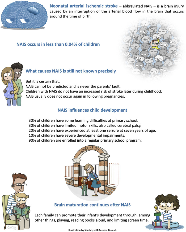
A new systematic review of the outcomes of neonatal stroke (Giraud A, et al. Long-term developmental condition following neonatal arterial ischemic stroke: A systematic review. Arch Pediatr. 2023). points out the uncertainties of prognostication in those babies also. They include this figure, as a suggested tool for counseling parents about prognosis.

There is a lot to like about this figure, especially the last section introducing things parents can do to promote development, but I am unsure about the reliance on percentages. Many people, physicians included, don’t understand what percentages like those mean for an individual child. There are studies to show that rates of outcomes are better understood than proportions. It is probably generally better to talk about how many children, out of a hundred children with a stroke, will have no learning difficulties in primary school, and how many will have difficulties.

The review article points out the high frequency of neurological or developmental concerns, but in fact most of the babies in the cohorts were functioning well. The first 4 lines on that figure about child development are all about the negative outcomes, even though they are a minority. Why not state them as positives?

“Of 100 children who had a brain problem like your child, 90 of them will go to normal school, but 10 will need special help with schooling. 70 out of every 100 children will do well at school, but 30 will have learning difficulties” might be easier to understand and focuses on the positive outcomes, experienced by the majority of children.

The plasticity of the neonatal brain makes our job, as prognosticators, more difficult and more uncertain than at any other age. Much of what is important in long term outcomes is invisible on brain imaging. From details of brain interconnections, to the rewiring of damaged regions, to family connections, parental interactions, how many books the family owns, attitudes to impairment, and future educational improvements; indeed all of the environmental influences on outcomes, that we can know little about in the NICU.

It is vital that we learn more about those other influences on outcomes and how to use them, but prognostication will always be uncertain, especially in the neonatal period, and even more so in the first few days after birth. We must be honest and transparent, and recognize and express the uncertainties with parents, while never minimizing their hope.

There is always room for hope, which may need to be adjusted but never destroyed. Hope in parents is associated with improved quality of life (Nordheim T, et al. Hope in Parents of Very-Low Birth Weight Infants and its Association with Parenting Stress and Quality of Life. J Pediatr Nurs. 2018;38:e53-e8), and parental peer support groups seem to enhance hope among participants (Dahan S, et al. Community, hope and resilience: parental perspectives on peer-support in Neonatology. J Pediatr. 2021;243:85-90 e2). Shared decision-making is enhanced when caregivers and parents share hope (Koch A, et al. Crossroads of parental decision making: Intersections of hope, communication, relationships, and emotions. Journal of child health care. 2023;27(2):300-15).

In “Candide”, Voltaire’s character Pangloss is a tutor of philosophy who believed that “all is for the best in this best of all possible worlds”, despite the evidence all around him, of the Lisbon earthquake which killed tens of thousands, and the Seven Years War which was raging. We must, in contrast remain reasonable and aware of the difficulties of some of those with major brain injury. We can express and inform parents about the range of possible, and likely, outcomes after a brain injury, while also recognizing the uncertainty of the future.

Posted in Neonatal Research | Tagged , , | Leave a comment

Closing the PDA… or leaving it alone?

Two new publications, in the Journal of Pediatrics, report trends in definitive closure of the PDA. This is a subject that I haven’t written about much on the blog, I wrote about the Beneductus trial last year, which was an RCT of early ibuprofen treatment of large PDA between 24 and 72 hours of age, compared to expectant management, without cox inhibitors, but with eventual surgical closure if needed. But I haven’t posted often about the PDA, and never, I think, about surgical/catheter closure.

You can see from the CNN 2021 report that there has been a dramatic reduction in definitive closure of the PDA (with surgery or a “device”) across Canada over the previous 11 years.

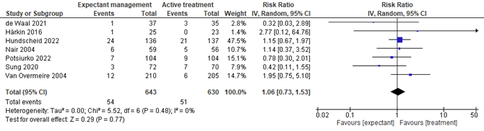
I guess for me the subject is murky, but as far as I can see through the murk, there is no clear indication for closing any PDA in the preterm. Outcomes between active treatment approaches and very restrictive approaches are quite similar. There is a very recent systematic review (Cheema HA, et al. Expectant management of patent ductus arteriosus for preterm infants: a meta-analysis of randomized controlled trials. Am Heart J. 2023) that examined clinical outcomes in trials which compared active therapy to trying to leave the PDA alone. They arbitrarily decided to include only trials with <25% rate of active treatment in the control group, I don’t know why 25% was the magic number. They showed no benefit of active treatment on mortality (all cause) or NEC, these 2 Forest plots are of mortality, and then NEC. (The figures are copied from the pre-proof, hence the watermark, or from the on-line supplement).

They found 7 trials that fit their criteria, 4 of them had very low rates of treatment among controls, between 0.7% (the Beneductus trial) and 8%. One of the trials was an RCT of prophylactic indomethacin, without confirmation of the presence of a PDA, so it was not an RCT of different approaches to the PDA.They also (Nair 2004) did not report the rate of treatment in controls, and it should really not have been included in this SR. Van Overmeire’s trial had a 24.8% rate of control treatment with cox inhibitors.

The review shows an increase in BPD with active treatment, as this Forest plot shows

The 95% confidence intervals cross the line of no difference, however, and if you delete the trial from Nair, with unknown control treatment rate, and unknown PDA status at the start of the trial, and Van Overmeire, with 25% treatment of the controls, then there is probably not likely to be a major impact on BPD one way or the other, but it seems that there is unlikely to be an improvement in BPD.

The following data from Pediatrix, (Shah ZS, et al. Trends in Procedural Closure of the Patent Ductus Arteriosus among Infants Born at 22 to 30 Weeks’ Gestation. J Pediatr. 2023:113716) among babies of <30 weeks gestation show a similar trend to the CNN, and they show the proportion with device closure, which has become more frequent than surgery. In the CNN, babies between 27 to 29 weeks now have an overall incidence of definitive closure of <1%, so a similar rate of definitive closure, and a major change in clinical practice over 10 years or less.

The other publication is from the Children’s Hospital Association in the USA, (Lai KC, et al. Current Trends in Invasive Closure of Patent Ductus Arteriosus in Very Low Birth Weight Infants in US Children’s Hospitals, 2016-2021. J Pediatr. 2023:113712) and only includes very preterm babies (<32 weeks) who were admitted for definitive PDA closure to a children’s hospital that was submitting data, between 2016 and 2021. The first surprise to me in this paper is that the number of admitted babies was relatively stable. I wonder where they are all coming from! As you can see from the part of the table that I reproduce below, apart from 2016, the numbers haven’t been going down very much, compared to the 2 other sources of data I have discussed. Of course, the ascertainment is completely different, but I would have expected a bigger drop in overall numbers, maybe referral patterns have changed.

They do, however, note the dramatic shift from surgery to catheter occlusion. They also note that the catheter procedures are being performed progressively earlier, decreasing from a median of 38 weeks PMA to about 31 weeks, and now similar to the age of surgery.


This dramatic change has taken place without any robust evidence that catheter occlusion is preferable to surgery. Or even much data that it is equivalent. I understand the desire to avoid a thoracotomy, but what is the relative impact of the 2 approaches on clinical outcomes? What is the relative effect on vocal cord paralysis and on vascular compromise? I presume that vocal cord paralysis would be much less frequent with catheter closure, and that vascular compromise of the limb that was used for the catheter would be much more common, but this incredible shift in practice, that has occurred without good studies, is worrying. The meta-analysis that was published a couple of years ago included only observational studies, there were no RCTs included comparing catheter closure to surgery. Its interesting also that the prominent, non-randomized trial of the Amplatzer device (Sathanandam SK, et al. Amplatzer Piccolo Occluder clinical trial for percutaneous closure of the patent ductus arteriosus in patients >/=700 grams. Catheter Cardiovasc Interv. 2020;96(6):1266-76) has the longest list of conflicts of interest that I can remember in neonatology:

S. Sathanandam: proctor/consultant Abbott; D. Gutfinger: full‐time employee Abbott; L. O’Brien: full‐time Abbott employee; T. Forbes: proctor/consultant Abbott, Edwards, AcuNav/Biosence Webster, B. Braun Medical, Siemens, Medtronic; M. Gillespie: proctor/consultant Abbott; D. Berman: proctor/consultant Abbott, Edwards, Medtronic; A. Armstrong: proctor/consultant Abbott, Edwards, Medtronic, B. Braun; S. Shahanavaz: proctor Abbott, Medtronic, and Edwards; T. Jones: research grant, proctor/consultant Abbott, Edwards, Medtronic, W.L. Gore & Assoc.; B. Morray: Consultant Medtronic, proctor Abbott; T. Rockefeller: proctor Abbott; H. Justino: proctor/consultant Abbott, Edwards Lifesciences, Medtronic; Clinical trial executive committee Janssen Pharmaceutical; Co‐founder PolyVascular; scientific advisory board Pediastent; D. Nykanen: proctor Abbott, consultant and independent data reviewer W.L. Gore & Assoc, expert witness Glaxo Smith Kline; E. Zahn: consultant/proctor Abbott, Edwards, Medtronic, National PI ADO II AS IDE Trial and Alterra/S3.

The 2 new publications give some data about outcomes: in the Children’s Hospital Association study the incidence of reported arterial thrombosis in the surgical closure group was 1.6% and venous thrombosis was 6%, with 3.6% being anticoagulated after the procedure. In the catheter occlusion group the arterial thrombosis incidence was 3.6%, and 4.4% had venous thrombosis, with 4.8% receiving enoxaparin. As the data are from an administrative database it isn’t clear if the thromboses occurred before or after the procedure, I presume they are retrieved from the recorded list of diagnoses during the hospitalisation. With the same limitations (and without necessarily routine surveillance) vocal cord paralysis was recorded in 4.8% of the surgical and 0.5% of the catheter closure group. There was less opioid use post-op in the catheter group (52% vs 87%).

The Pediatrix group study shows similar mortality in the surgery group to the catheter closure group, and a slightly shorter length of hospital stay (103 vs 109 days). It is hard to know what to make of length of stay in the other study, as many babies were transferred back to their hospital of origin.

I remain uncertain about the indications for definitive closure of the PDA. Most babies, even with a persistent shunt, can be weaned from respiratory support, and sent home, and most will eventually close spontaneously, although you might have to wait a few years. (Nielsen MR, et al. The chance of spontaneous patent ductus arteriosus closure in preterm infants born before 32 weeks of gestation is high and continues to increase until 5 years of follow-up. Acta Paediatr. 2022;111(12):2322-30). If you do need definitive closure, catheter occlusion looks like a reasonable alternative, and may be preferable, but it would be nice to have better data to confirm that.

Posted in Neonatal Research | Tagged , , , , | 2 Comments

Artificial Formulae and NEC. It is not necessarily the protein source!

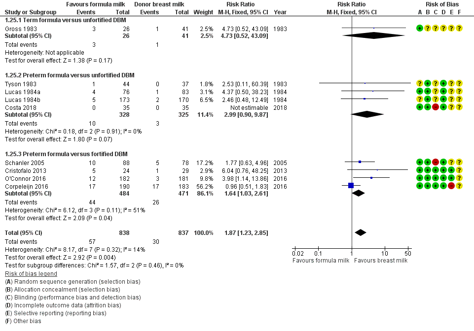
As mentioned several times recently, artificial formula seems to lead to an increase in Necrotising Enterocolitis, compared to donor milk. Of note, the trials in the Cochrane Systematic Review include both those with the entire diet being formula or donor breast milk, as well as those where the infants were randomized to formula or donor milk as a supplement to their own mother’s milk.

In the Forest plot from the Cochrane review, 3 of the 4 trials of “preterm formula versus fortified DBM” were among infants who had mother’s milk as the main diet, the entry criteria for Schanler 2005, Copeleijn 2016 and O’Connor 2016 included “mothers intended to breast feed”. The Corpelijn trial, however, only changed the diet for the first 10 days of hospitalisation, during which any supplement to mother’s own milk was either formula or un-fortified donor breast milk. In fact, I think there is an error in the classification of this trial in the Cochrane review, as far as I can see the babies in Corpelijn never received fortified donor milk; the babies received unfortified mother’s milk, which was supplemented either with artificial formula or unfortified donor milk until 10 days of age, after which they received maternal milk, with fortification according to local practice, and artificial formula as a supplement. The Cristofalo 2013 study only included babies whose mothers were not going to breast feed. Costa 2018 compared donor milk to artificial formula, but seems to have included babies whose mothers were not intending to breast feed, as well as those with insufficient milk. As a small study with no cases of NEC it is not informative for the NEC outcome in any case.

It is also useful to remember that the Schanler study had an extremely high frequency of NEC in the controls, and had nutritional practices which are not consistent with current best practice (“Administration of small quantities of MM (20 mL/kg per day) was initiated in the first week after birth and continued for 3 to 5 days before the volume was advanced. Milk intake was increased by 20 mL/kg daily to 100 mL/kg”), and it took an average of 18 days for the babies to receive even 50 mL/kg/day of milk. In contrast, in our NICU, similar babies of <1300 g (mean GA 27 semaines) achieved 150 mL/kg/day on average on day 16 with the previous version of our feeding protocol (it occurs now, with our progressive improvements in our feeding protocol, a little earlier).

Also, the Schanler 2005 study appears to have crossed over babies, and not analyzed them according to Intention to Treat: the methods include this phrase “Final group determination was made at the end of the study, on the basis of whether the participants had received the assigned supplement (Donor Milk or Preterm Formula)”. Which is very concerning, and raises doubts as to the reliability of the results.

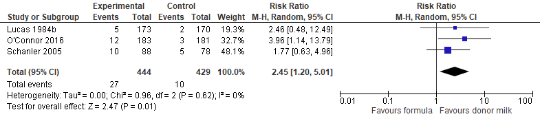
In order to show whether there is an impact on NEC, therefore, you have to include data from the much older studies of Lucas. The reports of those trials are often a bit confusing as there was more than one trial running simultaneously, and the results are partly pooled, they included babies up to 36 weeks gestation, and had an enormous incidence of NEC among infants of 34 to 36 weeks gestation (9% if they received only formula, and 0/113 if they received at least some breast milk). It is unclear to me how relevant these data are to current practice, but one of those simultaneous trials was investigating donor milk or formula as a supplement to maternal breast milk. It was included in the Cochrane review as Lucas 1984b, which is the term I use in the figure below.

This is a Forest plot of the effects of supplementation of mother’s milk with donor human milk (Experimental) compared to artificial formula (Control) on the incidence of confirmed NEC. Lucas 1984b did not use the classification of Bell, but their “confirmed NEC” is similar to Bell stage 2 or 3.

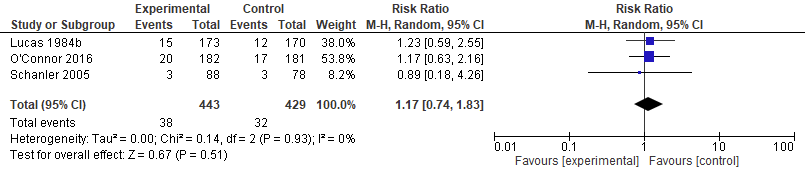
If we therefore meta-analyze those trials where babies seem to have received mother’s milk, and to have had a supplement of either donor milk, or artificial formula, and had their diet during the period of risk for NEC, we are left with just 2 fairly recent trials, and 1 much older trial, with the limitations noted above. This seems to confirm that artificial formula, as a supplement when there is insufficient mother’s milk, leads to a major increase in NEC compared to using donor human milk. Although the 95% confidence intervals are large, they do not include no difference, and all 3 of the trials are in the same direction with an I2 of 0. As you can see below there is no evidence of an impact on mortality, but the very small difference is in favour of donor milk.

There is no prima facie reason to think that the source of the proteins is the reason for the association between artificial formula and NEC. There are many differences between artificial formulas, pasteurized donor milk and preterm mother’s milk: the microbiome of the milk, the presence of Oligosaccharides (HMOs), the precise nature of those HMOs, the presence and concentration of intact human lactoferrin and immunoglobulins, the concentrations of insulin and leptin, and other components of milk are different between fresh maternal milk, pasteurized donor milk, and artificial formula.

So what is it about artificial formulas that leads to increased NEC? Is it related to disturbances in the intestinal microbiome?

A recent publication has reviewed the impacts of some food additives on the intestinal microbiome (Bancil AS, et al. Food Additive Emulsifiers and Their Impact on Gut Microbiome, Permeability, and Inflammation: Mechanistic Insights in Inflammatory Bowel Disease. J Crohns Colitis. 2021;15(6):1068-79). They review the extensive evidence, previously unknown to me, that a group of food additives, emulsifying agents, have been shown to have major impacts on the intestinal microbiome.

One of the emulsifiers that has an extensive record of altering the microbiome, and even being used to induce a model of ulcerative colitis in animals is Carrageenan, There is even a very recent review article describing all the adverse imapcts of Carrageenan, and how it causes colitis (Guo J, et al. How does carrageenan cause colitis? A review. Carbohydr Polym. 2023;302:120374). I was surprised, and disturbed, to see that Carrageenan is present in Similac special care 24 calorie milk. It is also present in Similac liquid fortifier, but not their powdered fortifier or the liquid fortifier with protein hydrolysate, nor indeed in Enfamil preterm formula, or Enfamil powdered or liquid fortifiers. They all also contain other ingredients, however, such as Soy Lecithin, and oils from various sources, used to provide DHA or ARA.

Any of the components of the artificial formulas that we give to preterm babies might have effects on their microbiome, or other effects on inflammation, or on the Toll-like receptors that are involved in the pathogenesis of NEC. Of note the preterm formula used in the 3 studies that I meta-analyzed above were not all the same, and indeed, the composition of the formulas may have changed between those publications and their current composition. The process of pasteurization induces changes in milk protein structure (including pasteurization of human milk, Sergius-Ronot M, et al. Impact of holder, high temperature short time and high hydrostatic pressure pasteurization methods on protein structure and aggregation in a human milk protein concentrate. Food Chem. 2022;374:131808.)

The take-home message: avoid artificial infant formulas during the period of risk for Necrotising Enterocolits. We don’t know why, but supplementing mother’s milk with artificial formula increases the risk compared to supplementing with donor human milk.

Posted in Neonatal Research | Tagged , , , , , | Leave a comment

Preventing NEC. Does erythropoietin have a role? Unreliable data.

I wrote a similarly titled post 3 years ago, which lamented the poor research practices of a group in Zhengzhou, who seem to perform large RCTs, then register them after completion, and sometimes register them with different primary outcomes to those which are then published. They often publish the articles in strange journals that rarely publish large clinical RCTs, journals which are often supposed to have editorial practices which do not allow the publication of retrospectively registered RCTs, but which publish them nonetheless.

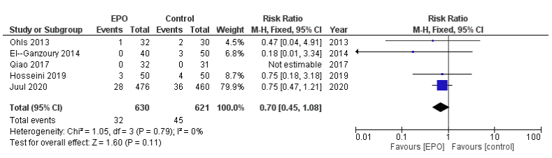
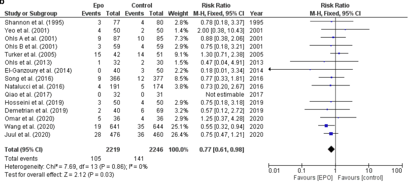
I bring this up now because of my series of posts on NEC prevention, and the publication last year of a systematic review of erythropoietin prophylaxis for preventing NEC. (Ananthan A, et al. Early erythropoietin for preventing necrotizing enterocolitis in preterm neonates – an updated meta-analysis. Eur J Pediatr. 2022;181(5):1821-33). That review showed, when including all the published data, a major, 23%, reduction in NEC with erythropoietin prophylaxis. However, when eliminating the retrospectively registered trials, there was no longer a clear effect, although a 50% reduction would still be within the confidence intervals of the meta-analysis.

Here is their Forest plot, from the on-line supplement, of the effects of prophylactic Epo vs control on the incidence of “definite NEC” that is grade 2 or 3, only including the prospectively registered trials

And here is their plot including all the RCTs

As you can see, the only individual trial which shows a reduction in NEC which is not likely due to chance is Wang et al, who had a very high incidence of NEC among their babies, with a mean GA of 30 weeks the controls had 17% NEC, with 5.4% stage 2 and 3 NEC. Among their subgroup of <28 weeks gestation 17% had grade 2 or 3 NEC in the controls. In comparison Juul et al report a control incidence of NEC of 8% among infants with a mean GA of 26 weeks.

Since that previous post the same group has published another retrospectively registered trial from some of the same institutions, with overlapping dates and overlapping eligibility criteria to the “epo for prevention of NEC trial”, the new trial is Song et al 2021, with the reference details below.

I wrote to the editors of the 2 journals which published the articles referenced below, pointing out the overlaps. This is an extract of that email:

Song J, Wang Y, Xu F, Sun H, Zhang X, Xia L, et al. Erythropoietin Improves Poor Outcomes in Preterm Infants with Intraventricular Hemorrhage. CNS Drugs. 2021;35(6):681-90.

Wang Y, Song J, Sun H, Xu F, Li K, Nie C, et al. Erythropoietin prevents necrotizing enterocolitis in very preterm infants: a randomized controlled trial. J Transl Med. 2020;18(1):308.

The two trials were performed by researchers mostly based in Zhengzhou, who have previously published an article on a related subject which was performed in 2009 to 2013 and retrospectively registered in January 2014 (Song J, Sun H, Xu F, Kang W, Gao L, Guo J, et al. Recombinant human erythropoietin improves neurological outcomes in very preterm infants. Ann Neurol. 2016;80(1):24-34). NCT02036073.

The two new trials enrolled babies of less than or equal to 32 weeks gestation, between January 2014 and December 2017 in the case of Song et al “in the neonatal intensive care unit (NICU) of the Third Affiliated Hospital and Children’s Hospital of Zhengzhou University”, and between January 2014 and June 2017 for Wang et al in “four centers including the Third Affiliated Hospital, Children’s Hospital, the First Affiliated Hospital of Zhengzhou University, and the Women and Children Health Care Center of Luoyang”. 

As far as I can see, therefore, there are 2 NICUs, at the Third Affiliated Hospital in Zhengzhou, and the Children’s Hospital of Zhengzhou, which were recruiting to both trials over the same period.

The eligibility criteria for the 2 trials were similar except with regard to the results of head ultrasounds performed before 72 hours of age.  In Song et al, only infants with Intraventricular Hemorrhage, of any grade, were included. In Wang et al infants with the more severe grades of hemorrhage were excluded, that is grade 3 and 4 hemorrhage. Thus, infants with grades 1 and 2 hemorrhage would have been eligible for both trials.

The CONSORT flow chart for Song et al notes that there were 370 infants with IVH admitted to the 2 enrolling NICUs during the period of the study, of whom 316 were randomized. According to table 4 in that publication there were 20 infants with the more severe grades of hemorrhage included in the trial.

The CONSORT flow chart for Wang et al reports that of 1327 infants assessed for eligibility, only 9 did not meet inclusion criteria (which should, therefore, include any babies with grade 3 and 4 hemorrhage) and there were 1285 babies randomized. There should therefore, according to Wang et al be a maximum of 9 infants with grade 3 and 4 hemorrhage across the four NICUs, but according to Song et al there were 20 in two of the NICUs.

There appear to have been 296 infants admitted to an NICU at the two hospitals involved in Song et al’s trial with grade 1 and 2 hemorrhage during the period January 2014 to June 2019. Over the same period 1285 infants were enrolled in those 2 hospitals and 2 other hospitals in a completely different trial with a different primary outcome. The 296 infants in Song’s trial would have been eligible for the Wang et al trial but are not mentioned in the Wang et al manuscript nor in their CONSORT flow chart.

Both trials were retrospectively registered after completion; NCT03914690 and NCT03919500 were registered within 2 days of each other in April 2019.

I believe that my observations raise serious questions about the research design, research ethics and publication ethics behind these publications. It seems that either there were substantial numbers of infants enrolled in both of the trials, or the CONSORT flow diagrams are inaccurate; there are, of course, other potential explanations.

Additionally, there are major problems with reporting within each of these manuscripts.

Song et al report none of the short-term outcomes reported as a routine in neonatal trials involving very preterm infants. They report a mortality of 25 infants of the 316 enrolled, a survival of over 92% of a group of infants with a mean gestational age of 30 weeks is remarkable.

The authors do not report the frequencies of bronchopulmonary dysplasia, retinopathy of prematurity, later serious brain injury on ultrasound, necrotising enterocolitis or late onset sepsis, all of which have major impacts on long term developmental outcomes. In order to determine the potential impacts of this prophylaxis in another group of very preterm infants, data regarding those other diagnoses is essential and should have been included in this manuscript.

The editor-in-chief of CNS drugs wrote back a few days ago, Sue Pochon is an employee of Springer Nature who appears to have no medical training, indeed her Linked_In profile shows that until 2001 she was catering manager at the Pirate Inn. She may well be an excellent manager, but I wonder if she has any idea of the importance of large RCTs in preterm infant showing an apparent major impact on NEC, and enormous apparent impacts on the developmental outcomes of the babies. The journal “CNS drugs” publishes a small number of articles each month, 6 or 7 usually, the large majority of which seem to be review articles, some systematic reviews, and a few observational studies. With a brief search I wasn’t able to find any other large RCTs, just one or two pilot trials. Unfortunately, their “instructions to authors” shows that they will accept retrospectively registered trials.

In her reply to me she notes that there was an investigation of my concerns and states “Fortunately, we have been unable to identify any fraudulent activity. While the trial dates do indeed overlap, we are satisfied that the trial participants were different, as were the trial outcomes, and that such concurrent studies are entirely feasible given the size of the recruiting hospitals.”

I had not, in fact, accused the authors of fraudulent activity. Rather that there were serious concerns, and that the CONSORT diagrams cannot be accurate. Either there were some babies whose data is included in both trial reports, or there are babies who were ineligible for one of the trials because they were enrolled in the other. Of note, the intervention in the 2 trials was identical (500 IU EPO/kg i.v. every 48 hours for 2 weeks) and controls received a saline placebo, even though they were not blinded studies.

The newer publication (Song et al, CNS drugs 2021) does include a very brief mention of some other clinical outcomes, such as a few cases of NEC in their babies, without specifying the grade of NEC, 11/159 [6.9%] in controls, vs. 9/157 [5.7%] in Epo babies. They also mention ROP being slightly lower, without specifying what stage, and BPD being significantly lower, without referring to any definition. According to the results of that trial, there was a dramatic effect on low Bayley (version 2) scores, with a more than 50% reduction in the proportion of babies with MDI <70, from 15% to 7%. They also show less CP, a reduction from 6% to 3%.

The whole point of registering a trial is to ensure that the sample size was calculated beforehand, that the primary, and main secondary, outcomes were decided before the data accumulated, and that the procedures were protocolized prior to performing the trial. With retrospective registration all this is lost. There is a risk that the primary outcome was chosen after examining the data, and/or that the study was terminated early when a potentially spurious outcome appears different between groups; both of which dramatically increase the chances of a type 1 error. These researchers have clearly been aware of the necessity of registering trials for many years, so they cannot even claim ignorance.

Journals should stop accepting retrospectively registered trials, they are unreliable sources of data that skew the medical literature.

The systematic review of erythropoietin for NEC prevention (referred to above) shows that it is indeed possible that Epo decreases NEC; the confidence intervals, even when the retrospectively registered trials are excluded, include a possible major reduction in NEC of 50%, largely based on the 25% reduction in grade 2 to 3 NEC shown by PENUT. This suggests to me that we need another large trial, focusing on NEC reduction, of prophylactic Epo. Apart from gestational age, there are few additional risk factors for NEC; early onset sepsis being one, but I don’t think you could do a trial just enrolling babies with EOS.

Any future RCT of NEC prophylaxis must be prospectively registered, with clearly defined primary outcomes, and eligibility criteria, and a report of the desired sample size.

Posted in Neonatal Research | Tagged , , , | Leave a comment

Preventing NEC : Gastric acid has a purpose, don’t block it.

As this series of posts have all had something to do with prevention of Necrotising Enterocolitis, I thought I’d write about some recent articles which refer to one of the risk factors for NEC, the use of acid suppression medications.

I’ve written previously about the risks of proton pump inhibitors (ppi) in young infants. They are a group of medications (along with Histamine receptor blockers) that I almost never prescribe. The commonest reason given for prescribing them seems to be gastro-oesphageal reflux, but of course they have no effect on reflux, just on the pH of the refluxed liquid, which is not necessarily a beneficial effect. If a baby had acidic reflux oesophagitis then maybe blocking acid production would be beneficial, but most reflux in newborn infants is non-acid, either neutral or alkaline. In fact, at least one animal study shows that ppis cause relaxation of the lower oesophageal sphincter, which might increase the risk of reflux!

A number of recent epidemiologic publications point out the complications associated with altering the gastro-intestinal physiology of a large number of babies. And the numbers are indeed large, prevalence of use in the NICU is not clear, but in France and in Scandinavia a substantial, and increasing, proportion of all young infants are exposed to ppi medications, somewhere between 2 and 5% of all infants!

We also should remember that we are really poor at diagnosing reflux. The only reliable way to know if a baby has an abnormal frequency of reflux is by impedance oesophageal recordings (multiple intraluminal impedance, mii), and a new publication has, yet again, shown that there is no correlation between a baby being irritable, or having arching episodes, and the presence of reflux events. (Njeh M, et al. The Irritable Infant in the Neonatal Intensive Care Unit: Risk Factors and Biomarkers of Gastroesophageal Reflux Disease. J Pediatr. 2023:113760). In this retrospective study they report over 500 babies who had pH-mii studies, and over 40,000 GER events with nearly 40,000 arching or irritability events. Their conclusion is quite clear ;

Acid GER disease is unlikely the primary cause of arching/irritability and empiric treatment should not be used when arching/irritability is present. Prematurity and neurological impairment may be more likely the cause of the arching/irritability. Arching/irritability may not be a concern in orally feeding infants.

So what are the complications of ppi use? The most immediately important in the NICU population is an increased risk of sepsis. This is true in older infants in the community (Lassalle M, et al. Proton Pump Inhibitor Use and Risk of Serious Infections in Young Children. JAMA Pediatr. 2023), in a nation-wide cohort study from France. It is even more striking among children in the PICU (Goyer I, et al. Proton Pump Inhibitor Use and Associated Infectious Complications in the PICU: Propensity Score Matching Analysis. Pediatr Crit Care Med. 2022;23(12):e590-e4), in this propensity score matched analysis, among children in the PICU, in Caen in Normandy,, who mostly received the ppi for stress ulcer prophylaxis, the risk of nosocomial infections was NINE times higher if they had received a ppi.

The other complication which has been confirmed in a recent study is an increase in the risk of fractures, this study is a propensity matched national epidemiologic study from the USA (Achler T, et al. Association of early-life exposure to acid-suppressive therapy and fractures during childhood: a retrospective cohort study. Arch Dis Child. 2023). This is probably due to effects of acid suppression on calcium absorption, which have been shown to increase osteoporosis risk in the adult.

Other studies from the last few years show an association of ppi use with the development of asthma, and with subsequent development of Coeliac disease, and the development of a variety of allergic diseases.

To return to the title of the post, does acid suppression increase the risk of NEC? PPI medications cause major changes to the intestinal microbiome (Levy EI, et al. The effects of proton pump inhibitors on the microbiome in young children. Acta Paediatr. 2020), and have been associated with LOS and NEC (See the following studies Manzoni P, et al. Exposure to Gastric Acid Inhibitors Increases the Risk of Infection, Patil UP, et al. Efficacy of and potential morbidities associated with the use of antacid medications, and this review article (Tan J, et al. A Review of Histamine-2 Receptor Antagonist and Proton Pump Inhibitor Therapy for Gastroesophageal Reflux Disease in Neonates and Infants. Paediatr Drugs. 2023;25(5):557-76). As there are almost no RCTs of these agents in the preterm infant, there is no reliable evidence of the size of the risk, but as that review article points out there is no evidence of any benefit either. The epidemiologic studies suggest that there is no benefit, including among babies with a clinical diagnosis of reflux.

One situation in which a ppi is frequently prescribed is after Gastro-oesophageal fistula repair. Oesphageal dysmotility is universal after such surgery, and there is a risk of developing anastomotic strictures. A ppi or another antacid medication are often prescribed after surgery, just in case the infant has acid reflux, and in case the reflux might increase the risk of stricture. A recent publication from my centre (Righini Grunder F, et al. Should Proton Pump Inhibitors be Systematically Prescribed in Patients With Esophageal Atresia After Surgical Repair? J Pediatr Gastroenterol Nutr. 2019;69(1):45-51) shows that routine prescription of a ppi does not prevent formation of strictures, and that many babies were probably receiving them for other symptoms, such as those due to tracheomalacia, for which they are probably ineffective.

A final concern, ppi use induces hyperplasia of the parietal, acid-producing cells, by a secondary increase in gastrin production. Which means that there is often HYPERacidity when then are eventually stopped. Hence the title of the article from which this image is taken “Evidence That Proton-Pump Inhibitor Therapy Induces the Symptoms it Is Used to Treat“. They note in that article that Histamine-2 receptor blockade does the same thing.

My take home message? Don’t.

Don’t prescribe acid suppression therapy for suspected reflux.

Don’t prescribe acid suppression therapy for proven reflux, unless you can show acid oesophagitis

Don’t prescribe acid suppression therapy for prophylaxis against upper GI bleeding.

Don’t prescribe acid suppression therapy for preterm infants at any risk of NEC.


Posted in Neonatal Research | Tagged , , , | 2 Comments

The myth of the “exclusive human milk diet”; fortification options for breast milk.

The Exclusive Human Milk Diet sounds superficially immediately convincing: “We should only give milk products derived from human milk to human babies”.

But, is it more than just a catchy phrase? The major company which is responsible for producing a human milk based breast milk fortifier is heavily into promoting its use. They are certainly responsible for substantial profits for their shareholders. Unfortunately, many of the publications about the EHMD have been marked by important conflicts of interest, and, most importantly, they have not performed the most important relevant comparison. Which is, if you only use human milk (mother’s or donor) for every feed of the very preterm baby, is there an difference in clinically important outcomes between a multi-component fortifier derived from donor human milk, and one derived from bovine milk?

What does the evidence currently show?

Mother’s own milk, unpasteurized, is almost certainly associated with the lowest rate of NEC compared to other sources of milk. If there is insufficient mother’s milk, then pasteurized donor milk is the next best choice, leading to lower rates of NEC than artificial formula. I think this is now well-enough established, despite the limitations in the data, that I won’t even put the references in.

When it comes to breast milk fortification, is there any evidence to support the use of human milk derived fortifier compared to bovine milk derived fortifier, among infants who only receive human milk feeds?

There are only 2 relevant trials, one I have discussed previously, more than once, from Toronto, O’Connor et al, which randomized infants, who were all receiving only human milk feeds, to a bovine fortifier or a human milk derived fortifier, and found no effect on NEC or other clinically important outcomes.

The other is a Swedish trial which is now available as a preprint. Jensen GB Effect Of Human Milk-Based Fortification in Extremely Preterm Infants Fed Exclusively with Breast Milk: A Randomised Controlled Trial. Available at https://papers.ssrn.com/sol3/papers.cfm?abstract_id=4529245. This is the not yet peer-reviewed report of the results of a trial for which the protocol has previously been published. (Jensen GB, et al. Nordic study on human milk fortification in extremely preterm infants: a randomised controlled trial-the N-forte trial. BMJ Open. 2021;11(11):e053400).

In this new trial 229 infants <28 weeks GA were randomized before they reached 100 mL/kg/d of oral milk feeding. They all received mother’s milk or donor human milk, and the milk fortification was individualized, with standard fortification plus extra protein or fat as required, all of them being from human milk in the intervention group, or cow’s milk in the controls. The cow’s milk fortifier used was not standard between the 7 NICUs involved, but they continued to use whatever they were previously using. The study babies were not allowed to receive formula in either group prior to 34 weeks PMA.

The primary outcome was a composite of death, NEC, and late-onset sepsis, prior to discharge. All x-rays were independently analysed, and outcomes were assigned by investigators masked to group participation.

There was no difference in the primary outcome, nor in any component of that composite, in particular there was no difference in Necrotising Enterocolitis.

You can see the results below:

If I do a quick and dirty meta-analysis of the 2 trials, assuming there are no other studies that I am unaware of (yes, it does happen!), we can see the following outcomes, using a random effects model, and the RevMan version 5 software:

Necrotising Enterocolitis, Bell stage 2 or 3

Mortality

Late-onset Sepsis

There is no clear difference in any outcome between the different sources of breast milk fortifier.

A recent article claimed to show that human milk derived fortifier was cost-effective. The costs of the human milk derived fortifier, among the 7 hospitals involved, were between about 250,000 and 1.6 million annually, and they claim that there was an overall cost saving associated with the EHMD (of supposedly up to 3.4 million dollars a year, a number which is being touted by the major producer in their publicity). However, that publication was based on no presented data, just what participants in a round-table discussion said to each other; I really don’t understand how it got published.

In fact if you do an internet search you may well see the headline “Prolacta fortifiers save hospitals up to 34M annually”! Clicking on the link will make it clear that is up to 3.4 million (not 34), but nowhere is it clear that the cost savings are only if you compare using Prolacta products to using formula as supplement to mother’s milk.

I think it is, indeed, likely that an EHMD diet might save money if you compare it to using artificial formula, but that is not a choice which any of us would now make. As Human milk derived fortifier is very expensive, at somewhere over $10,000 US per baby, and there is no clear advantage at all over bovine milk derived fortifier, then it can’t possibly be cost effective in comparison to using a cow’s milk based fortifier.

There are several review articles, often, again, marked by conflicts of interest, which are sometimes not clearly declared, which suggest that it is important to avoid bovine milk proteins, to reduce NEC. That assertion cannot be supported by the literature. There are many differences between human milk as currently used (either fresh mother’s milk, or pasteurized donor milk) and artificial formula; the extensive processing of bovine milk to create sterile formula, and the lack of Human Milk Oligosaccharides in the product, the lack of human immunoglobulins, and many other differences may be much more important than the source of the protein. Preterm babies are immunologically incompetent; there is no primary reason to suppose that artificial formulas increase NEC because of the source of the protein, it may well be because of some of the many other differences between formula and human milk.

Another issue is that breast milk has become a saleable commodity in some places, the New York Times has an interesting article discussing some of the implications of this, one of which is that poorer mothers could possibly end up selling their milk instead of giving it to their own babies. In most places outside of the USA breast milk donation is an altruistic act of lactating mothers, to whom we should all be immensely grateful.

To be strictly evidence-based, as I always strive to be, the current evidence (as you can see in the Forest plots above) continues to have wide confidence limits, and is therefore consistent with either a substantial increase or decrease in NEC with human fortifier compared to bovine. If the producers of human milk based breast milk fortifier want to continue to promote it as a way of reducing NEC, compared to a human milk diet fortified with bovine fortifier, then they should be forced to perform an adequately powered trial.

Even a 25% reduction in NEC would be of major clinical benefit, and a reduction from 8% to 6% might be cost effective too. It would also be within the confidence limits of the currently available data. Such a reduction could be sought with a RCT with a sample size of about 2,500 per group. If that was prohibitive, and I don’t think it should be given the enormous profits possible if they could prove efficacy for reducing NEC, then focusing on a higher risk group, such as the <26 week infant, and hypothesizing a larger decrease such as reduction of 40%, could give a more manageable sample size. A reduction from 10% to 6% would need a sample size of about 700 per group, with a power of 80% and an alpha of <0.05.

As the evidence currently stands, I consider the myth of the Exclusive Human Milk Diet : BUSTED.

Posted in Neonatal Research | Tagged , , , , , | 11 Comments

Necrotising Enterocolitis: impacts of feeding patterns

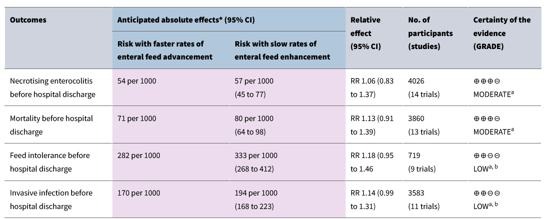
A new review article published in J Perinatol makes many claims about feeding patterns and NEC, but I think has seriously misinterpreted the literature (Assad M, et al. Dilemmas in establishing preterm enteral feeding: where do we start and how fast do we go? J Perinatol. 2023;43(9):1194-9). It is a little strange, that only 9 months ago a very similar publication, with a very similar title, appeared in the same journal, (Patel AL, Taylor SN. Dilemmas in initiation of very preterm infant enteral feeds-when, what, how? J Perinatol. 2023;43(1):108-13) and came to many different conclusions.

Trophic feeding

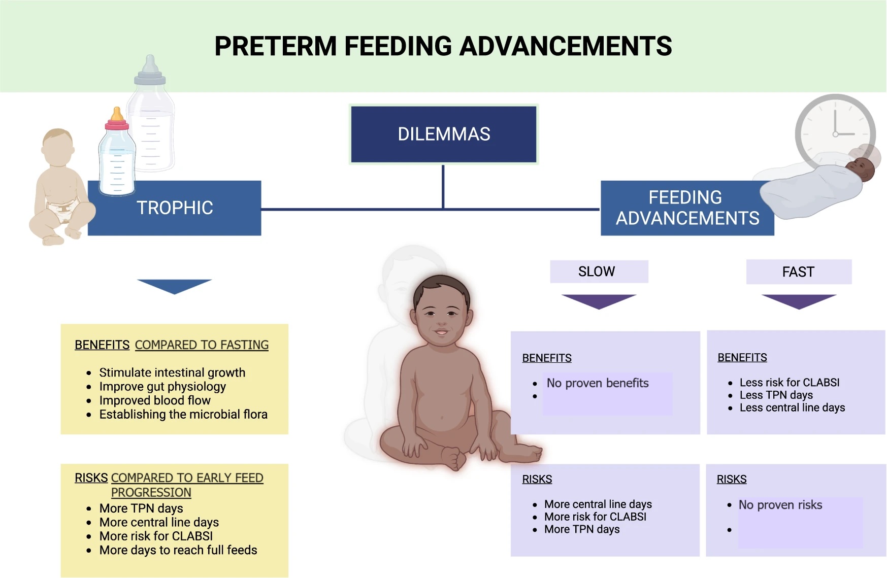
In the new article, the authors suggest that trophic feeding is advantageous, but that, of course, depends on what you compare it to. Trophic feedings are clearly preferable to a period of fasting, in terms of the physiologic responses, and do not increase (or decrease) NEC compared to a period of fasting. There is a Cochrane review, which dates from 2013, and showed no substantial benefit or risk of early trophic feeding, compared to a period of fasting: one further study, published since then, (Tewari et al 2018) also showed no adverse effect. In terms of limitations, few babies of high risk were included in those trials, and they tended to have very slow rates of feeding advancement once feeds were increased. Assad et al list the probable benefits of trophic feeding, but do not clarify that that is in comparison with a period of fasting.

Feeding patterns after a trophic period

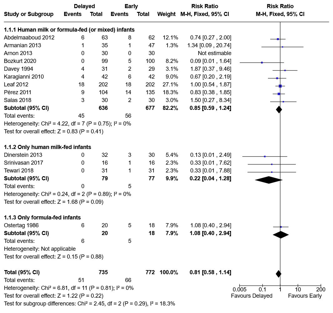
Once you start to feed after a period of trophic feeding, the new review contrasts the study by Berseth 2003 to the trial of Salas 2018, suggesting that the results are not consistent. It is not surprising that the results were not consistent, however, as the trials were vastly different. The comparison made by Berseth was a period of trophic feeds, compared to immediate feeding advancement, among infants who had been starved for an average of 9 days after birth, and sometimes as long as 31 days! Indeed, the only study of different feeding patterns, or rates of advancement, that has ever shown an impact on NEC is that study by Berseth. Which means that if you are going to starve your babies for a week, then a period of trophic feeds is probably a good idea before increasing them. But most of us don’t do that anymore (or never did!).

The Salas study, in contrast, shows that among babies who were npo for 1 to 3 days after birth, immediate advancement, compared to 4 days of trophic feeding, did not lead to more clinical problems. There isn’t much other data comparing a period of trophic feeding to immediate progressive feeding increases.

Early feeding advancement

There are several trials that have investigated the impacts of starting feeds early, to starting them later, without a period of trophic feeds, in other words with an immediate start of feeding advancement. One of the best was limited to only very high risk babies, with SGA and abnormal antenatal dopplers. This was the ADEPT trial (Leaf 2012 in the figure below), which showed that the babies in either group had a lot of difficulty with feeding tolerance, but that there was no difference in outcomes of NEC, or survival, by group assignment (starting enteral feedings and immediate advancement at 2 days compared to at 6 days.) The Cochrane review showed no impact of delayed progressive feeds, compared to early progressive feeds, the Forest plot below is for the outcome stage 2 NEC, and shows that earlier feeding does NOT increase NEC.

Speed of feeding advancement

Once you start feeds, then the rate of increase has NO impact on NEC from any of the randomized trials. That is the other very misleading statement in this new review, the figure and the text suggest that slower feeding advancement leads to “possibly less risk for NEC” and “less risk for feeding intolerance”. Neither statement is supported by the literature.

The Cochrane review shows no hint of an increase in NEC with more rapid feeding advancement, the actual proportions with NEC are very slightly lower among those with more rapid feed increases. Similarly, there is no evidence that slower feeding advancement improves feeding tolerance. Feeding intolerance is, of course, a very difficult thing to quantify, but this is certainly consistent with my observations, some babies have regurgitations during the first few days of feeding, no matter how quickly you aim to advance the feeds. The comparisons in the various RCTs have usually compared a slower rate of feeding advancement to 30 mL/kg/day, occasionally as fast as 35 to 40 mL/kg/day.

This new review article sees to be based on personal preferences and inbuilt prejudice, with no evidence-base for many of the statements made. The following feeding plan is based on the conclusions of my review of the literature, and is very similar to the conclusions of an excellent published review from 4 years ago, (Kwok TC, Dorling J, Gale C. Early enteral feeding in preterm infants. Semin Perinatol. 2019;43(7):151159):

A feeding plan consistent with the best evidence

Some of the evidence is admittedly limited for some comparisons, especially in the very highest risk populations. Babies in shock or on inotrope/vasopressors could be an exception to the following plan, and they could be either kept fasting, or on low volume trophic feeds, but there really is no evidence to decide how to feed such babies, who may have reduced intestinal blood flow, and a limited capacity to increase flow after feeding.

There is no evidence that prolonged fasting, several days of trophic feeds, or slower advancement of feeds have any benefit in terms of NEC or feeding intolerance. Arriving at full feeds more slowly, however, does increase the duration of TPN, and all the associated complications, including late-onset sepsis (although, to be as stringently evidence-based as possible, the evidence for a reduction in LOS with more rapid advancement is uncertain).

Enteral feeding should be started on the first day of life, with maternal breast milk if at all possible. Feeds should immediately be advanced, by at least 30 mL/kg/day if tolerated, to between 160 and 200 mL/kg/day.

That’s it. Simple really! Give it a try.

In the new publication, there is a cute figure which summarizes the misinformation, and which I have, unfortunately, seen shared approvingly. With apologies to the authors, who I’m sure have the best intentions, I have corrected the figure to make it evidence-based.

Posted in Neonatal Research | Tagged , , | 4 Comments