Probiotics might save lives in low-resource countries

After several years of preliminary investigations,  a huge RCT has been published from India (Panigrahi P, et al. A randomized synbiotic trial to prevent sepsis among infants in rural India. Nature. 2017;548:407.) which enrolled babies over 2 kg birth weight between 24 and 96 hours of age to receive a synbiotic mixture, Lactobacillus plantarum with a fructo-oligosaccharide, which they received for 7 days. This followed studies showing that this mixture led to stool colonization for several weeks of life. All the babies were breast-fed, although a few received additional liquids by mouth, including water and honey.

The babies were then followed for up to 60 days to see if they needed evaluation for sepsis, which was diagnosed by WHO criteria. The primary outcome of the trial was the combined outcome of death or sepsis.

They planned to enrol over 8,000 babies, but stopped early after “only” 4,556 babies for efficacy.

The combined outcome of death or sepsis was reduced from 9% in the controls to 5.4% in the synbiotic group. The entire difference being in sepsis, as mortality was low (<0.3%), there was a reduction in gram-negative sepsis, gram-positive sepsis and lower respiratory tract infections, as well as in culture-negative sepsis.

The study was a remarkable achievement, an individually randomized, placebo controlled trial in over 150 villages in India. The study excluded smaller babies, and those thought to be under 35 weeks, as well as 254 suspected to be septic at the time of screening for study entry, and those whose mothers appeared to have perinatal infections. The total number of exclusions came to 2,506. The synbiotics were supplied in capsules with “mixing containers and syringes with needles” initially kept at -20 degrees Celsius then distributed with cold packs. I am not sure exactly how the synbiotics were administered, which I think is an important detail for future wide implementation. I presume the syringe and needle were used to inject a solvent (?sterile water) into the capsules and then aspirate it prior to administering the liquid into the mouth of the baby.

The technical difficulties in distributing and administering this synbiotic preparation as a routine will need to be addressed, but this is an intervention with little or no risk that could improve outcomes for millions of babies around the world.

Posted in Neonatal Research | Leave a comment

Endotracheal intubation, making it safer for babies

Many of our patients need invasive ventilatory support, for which endotracheal intubation is required, but we intubate many fewer babies than in the past. We also very rarely intubate babies for endotracheal suction to remove meconium any longer. Which means that there are vastly fewer opportunities for trainees to learn intubation. I never kept a log while I was training but I am pretty sure that I intubated more babies in my first 2 weeks on a neonatal service than residents now do in their entire training. In addition to the above factors, extended role respiratory therapists and nurse practitioners also need training and experience.

Training juniors and other practitioners to intubate is something I have done many times, usually by showing them the appearances during a laryngoscopy, and talking through the procedure as I performed it, followed by close supervision for a few intubations. It is difficult however, sometimes, to see what they are doing wrong (except when they pick up the laryngoscope with the wrong hand, for example).

There are now 2 RCTs of the use of a video-laryngoscope during the training of intubators in the NICU (one of which was done in my institution, the PI being my colleague Ahmed Moussa), and 2 recent crossover trials of using the video-laryngoscope during training in the simulation lab with mannequins (one of which was by the same Dr Moussa Assaad MA, et al. Learning Neonatal Intubation Using the Videolaryngoscope: A Randomized Trial on Mannequins. Simul Healthc. 2016;11(3):190-3) this is the other one  Parmekar S, et al. Mind the gap: can videolaryngoscopy bridge the competency gap in neonatal endotracheal intubation among pediatric trainees? a randomized controlled study. J Perinatol. 2017.

The results are very consistent, trainees intubate with much more success during initial intubation attempt when using the video-scope, but they take a little longer per attempt; when they then attempt intubation with a conventional laryngoscope they maintain the skills that they learnt.

The two clinical trials also showed similar results, success on initial attempt was much higher with the video, this was despite difference in study design, in the study mentioned already the residents were randomized to using either the video-laryngoscope or a conventional scope, in the other trial they all used the video-scope but were randomized to having the screen covered or not the trainees did not look at the screen in this second study, the supervisor looked at the screen to give them guidance about their technique. The duration of intubation was a little longer with the video compared to the conventional, (Moussa et al) but identical between the video with screen visible and with screen hidden groups; O’Shea et al.

Why do trainees fail to intubate? I think we know the reasons why trainees fail to intubate, but the relative frequency of those causes, and how we can use video-laryngoscopes to reduce and correct them was, I think, unstudied. Until the remarkable Peter Davis (who seems to publish more clinically useful research than anyone else in neonatology, it is hard enough to keep up with the neonatal literature, Peter single-handedly makes it much harder!) and his group reported these data : O’Shea JE, et al. Analysis of unsuccessful intubations in neonates using videolaryngoscopy recordings. Archives of disease in childhood Fetal and neonatal edition. 2017. This is an analysis of video-recordings which were made of both groups in the above mentioned study, even the babies in the screen hidden groups had the videos recorded.

You can see here the categories of failure reasons in the 2 groups, which are similar apart from failure to recognize the vocal cords, presumably a supervisor watching the screen could say “look, there are the cords!” (to which the residents does not respond verbally, while thinking “oh, so that’s what they look like, why aren’t they yellow like in the text-book?”)

The degree of visualization of the glottis was analyzed, and the video-visible group (the intervention group) achieved better visualization, presumably because of the coaching.  (of note there is a header missing over the last column of this table in the published version of the article, I presume the header says ‘C-L grade when inserting ETT in intervention videos (n=14), n%):

I think the video-laryngoscope is clearly an essential tool for teaching neonatal endotracheal intubation, you have to be aware that there are differences between the available devices, the 2 clinical trials highlighted here used different scopes, we use the Storz device, which does not have a 00 blade, and has a somewhat bulkier blade design than a standard Miller blade. Because of this I don’t use it for intubation below about 750 grams. As we don’t teach intubations on babies under 29 weeks that doesn’t cause a huge problem, but I would like to be able to demonstrate the anatomy on smaller babies, and I think even for more senior trainees with experience in intubating bigger babies, the capacity to use the video-scope for coaching in the littlest ones would be invaluable. The Melbourne group used a “Lary-flex” from Acutronic, with a blade which looks more like a traditional Miller blade, and has a 00 available.

As they note in their article, blade design, even among blades which are all named “Miller” differs between manufacturers, a fact of which I have had personal experience, when a hospital changed suppliers, as the alternative was cheaper and had the same appellation, but the blades were quite different and much more of a problem for intubating tiny babies.

Should all intubations be performed with a video-laryngoscope? In the NICU there is little good evidence about use of the video other than for training. A brand new systematic review of intubation complications and how to reduce them (Cabrini L, et al. Tracheal intubation in critically ill patients: a comprehensive systematic review of randomized trials. Critical Care. 2018;22(1):6) showed no clear benefit of video-laryngoscopy for routine intubation, or high risk intubations, and even a higher risk of complications compared to standard laryngoscopy. That is based on post hoc analysis of data from the 2 largest trials (out of the 9 total trials that they found, all in adults).

A review in pediatric patients from a few days ago (Xue F-S, et al. Paediatric video laryngoscopy and airway management: What’s the clinical evidence? Anaesthesia Critical Care & Pain Medicine. 2018) found a large number of articles in children, most of which either excluded newborns, or included very few of them. They were unable to find convincing evidence of the benefit of the video-laryngoscope in clinical practice, and noted the great variations between the 5 models they reviewed (which did not include the Lary-flex).

I think trainees should be taught to intubate using the video-laryngoscope, and, until they are clearly highly competent, all their intubations in the NICU, and perhaps in the delivery room, should be supervised by someone who is highly competent and experienced, and is reviewing the screen showing a video of the process.

Introducing the video for routine intubation of the newborn by individuals who are already experienced and highly competent is not currently supported by any good evidence, although my feeling is that with optimised equipment it may one day become standard of care. This may need further refinement of the equipment, and improvement in blade design, including availability of 00 blades for the tiniest babies.

Endotracheal intubation is associated with frequent adverse events, some serious. Improving the safety of our patients, while ensuring the competence of our trainees as they prepare for independent practice is essential.

Posted in Neonatal Research | Tagged , | 1 Comment

Does tactile stimulation in the delivery room actually do anything?

One of the things that is done in neonatal resuscitation that isn’t part of resuscitating older patients is tactile stimulation. Babies who are apneic and/or floppy often receive stimulation in the form of rubbing the back, patting or flicking the feet, or, in the old days, slapping the butt (sometimes while held upside down by the feet!)

I have never been quite convinced of the efficacy of this, but haven’t thought much about how you would prove if it works or not, or even what “works” would mean. Does tactile stimulation actually start babies breathing sooner? Does it delay effective ventilation? In babies who are bradycardic is there any benefit?

I have seen babies receiving prolonged stimulation with little response, and have even been pushed aside by older nurses while I was trying to ventilate a baby so that they could give a really good rub of the babies back! It is such an ingrained practice that it is accepted uncritically in the initial steps of NRP.

I think it is possible that stimulation has no effect at all, and that the common observation of babies starting breathing after stimulation promotes a confirmation bias, “because they started breathing after being stimulated it must have been because of the stimulation”.

I think it is also possible that stimulation causes reflex respiratory effort, but only in babies that would have started breathing a few seconds later in any case, with no overall benefit.

I think it is also possible that there is enough impact that babies with moderate depression at birth might have a benefit, starting to breathe effectively earlier and even perhaps having an increasing heart rate if mildly bradycardic.

I think it is unlikely that a seriously depressed baby has a benefit, and that, for such a baby, stimulation runs a risk of delaying effective interventions.

How would you prove any of this? It would be difficult to do a prospective RCT, you would have to randomize babies to 2 different NRP type protocols, one with stimulation and one without, you’d need enough babies to include a substantial number who actually need intervention, and then you could look at time to effective ventilation, time to a stable heart rate, or something like that, as the outcome criterion. Could you ethically randomize babies to a no stimulation protocol? Could you get enough people to refrain from stimulation for the study to work?

I think the first thing should be to collect some reliable observational information about the impacts of stimulation. This new study has done just that: Baik-Schneditz N, et al. Tactile stimulation during neonatal transition and its effect on vital parameters in neonates during neonatal transition. Acta Paediatr. 2018 Video recordings and pulse oximeter recordings of babies during transition were analyzed. There were just over 50 term and 50 preterm babies included in the analysis. 18 of the preterms and 25 of the term babies were stimulated at some point. In the preterm group babies were often given respiratory assistance without stimulation, which rarely happened in the term babies, for whom most of the non-stimulated babies did not need any intervention. They weren’t able to measure direct respiratory impacts of stimulation, but they did show in the term babies that there was no change in the saturation or heart rate before and after the stimulation, (comparing the averages for 30 second periods). In the preterm babies heart rate did not change, but saturations increased (which of course might have happened anyway…).

Another study Dekker J, et al. Tactile Stimulation to Stimulate Spontaneous Breathing during Stabilization of Preterm Infants at Birth: A Retrospective Analysis. Frontiers in Pediatrics. 2017;5(61) analyzed practice regarding stimulation among preterm babies. The basic message is that practice was extremely variable. Which to my mind is completely appropriate, as there is really no evidence base for tactile stimulation at term,  and even less (if you can have less than zero without becoming negative) after preterm delivery, then variability in practice should be expected, and should give room for observational studies, and then prospective studies.

Millions of babies are born every year who receive some sort of intervention after birth, failure to adequately adapt to life outside of the womb is a major cause of death and long term disability in the world. Our review of standardized training programs showed that such programs seem to improve survival, and that most survivors have good outcomes.

While awaiting good prospective studies what should we do? I think that, for a term baby with a good heart rate who does not breathe quickly after birth, rubbing the back or flicking their feet is probably harmless and might stimulate an increase in respiratory drive. If the baby is bradycardic or fails to respond within a very short interval (perhaps 20 seconds) then tactile stimulation should quickly be abandoned in favor of positive pressure ventilation.

Posted in Neonatal Research | Tagged | 3 Comments

New Published Letter, a response to the ‘number needed to suffer’.

The new issue of Acta Paediatrica (Februray 2018) includes a letter I wrote in response to a commentary written by a paediatric anaesthetist, Dr Lönnqvist.

If you receive an email with a description of the contents of each issue, as I do, you may have seen this :

A number of papers explore the outcomes of extremely preterm infants. Gillone et al state that the one-year survival rate for babies born at less than 501 grams in the North of England was only 22% in 1998-2012. Lynoe et al ask what the indications should be for providing extremely preterm infants with neonatal intensive care and Lönnqvist proposes that disability-free survival and the number needed to suffer should be used as outcome measures for such care. He claims that it is misleading to just focus on saving lives, without paying attention to the risk of severe disabilities, poor quality of life and the suffering of families. Helgesson agrees with Lönnqvist, while Barrington strongly disagrees.

That brief description mischaracterizes the points that I made. Dr Lönnqvist wrote his ‘A Different View’ claiming that disability-free survival was a new concept that should be used as the primary focus on decision making for extremely preterm infants.

That, of course, is ridiculous, as I noted in my letter, disability-free survival has been an unfortunate focus of neonatal outcome studies for decades; I say unfortunate because calculation of such an index conflates death and ‘disability’. The point I made is that both death and disability are important, but they should be considered separately, not lumped together as if they had equivalent impact, or were treated and valued the same by families. Of course, as you will know if you are a frequent reader of this blog, so-called ‘disability’, as included in most long term outcome studies, usually means low scores on developmental screening tests, and not the presence of a disabling condition.

To emphasize a point I have made many times, “NDI”, which stands for Neuro-Developmental Impairment, is an abusive misnomer. Most babies who are thus labelled are listed as being impaired because of lower scores on developmental screening tests, like the Bayley Scales of Infant Development. But a Bayley version 2 MDI score under 70 (or a Bayley 3 language or cognitive composite under 85, or indeed a score on any standardized developmental screening test below an arbitrary cutoff) is not an impairment. An impairment is “a loss or abnormality of psychological, physiological or anatomical structure or function”; infants with a developmental test score below a particular threshold do not, by this definition, have an impairment. If their developmental difficulties affect their function, then they may have an impairment, but most babies with lower screening scores have no functional ‘abnormality’, and are not impaired.

Dr Lonnqvist’s belief that disability-free survival was a new concept demonstrated to me how little he knew about the field. But more disturbing to me was Dr Lönnqvist‘s attempt to create a new metric to determine the impact of survival among high-risk newborns. He called this the ‘number needed to suffer’, which is calculated as I noted in my reply, part of which is reproduced below:

Dr Lönnqvist’s calculation of the ‘number needed to suffer’ requires that he conflates impairment with suffering. His idea that a day of living with an impairment is equivalent to a day of suffering is completely unacceptable. His calculations of ‘number needed to suffer’ require that infants who die before they leave the NICU have the sum of their ‘days of suffering’ calculated. This is then added to the number of days of life of children at follow-up who have impairments to calculate the denominator of suffering. According to his calculations, the numerator is the number of babies who survive without impairment. Again, according to this nonsensical metric, as the children get older the ‘number needed to suffer’ increases each day, because, according to this calculation, having an impairment means daily suffering. At 3.5 years, he calculates a number needed to suffer of 1/1369, at 10 years, if we counted minutes rather than days, the NNS would be in the billions!

As I point out in my letter, an issue to which Dr Lönnqvist does not respond in his answer (also published on-line simultaneously), if we follow his reasoning, any surgery for a child who is already blind or has cerebral palsy would be unconscionable, their ‘disability free survival’ would be zero and their ‘number needed to suffer’ would be infinite.

Despite the prejudices of Dr Lönnqvist and others of like mind, most extremely preterm babies who survive do so without serious impairment, even those who really do have serious impairments have a good quality of life, and there is little impact of gestational age at birth on the frequency of impairment.

In his reply to my letter, Dr Lönnqvist suggests that babies in the south of Sweden born at 24 weeks gestation have better outcomes than babies in the North, and that this is because of the vacuuming up of resources by the 22 weekers, who are much more likely to receive active care in the North than in the South.

This is, of course, nonsense. Admitting one of the 3 or 4 babies of 22 or 23 weeks gestation each year in an NICU has no perceptible impact on resource utilisation overall for an NICU (there were about 50 deliveries at 22 weeks over a 3 year period, they were admitted to one of 7 NICUs). There is no reasonable way you could calculate the impact of such an admission. The data presented by Dr Lönnqvist are neither peer-reviewed nor are they adjusted for baseline risks, he presents unverifiable data (which incidentally appeared on the front cover of Acta Pediatrica a few years ago) from what he says are Swedish government sources (in the legend to the figure he states that these are from “post-publication data processing” of the EXPRESS cohort) we have no idea if those data include delivery room deaths, how they were collected etc. etc. In fact the numbers in that figure are not the same as any numbers in the original cohort, neither the numbers of fetuses alive at admission of the mother, nor the number of live births.

In the original publication prior to the “post-publication data processing” you can see from this figure that the births at 22, 23 and 24 weeks were considered together. You can also see that the centers with the lowest odds of death at 25 to 26 weeks were the centers that also had the lowest odds of death at 22 to 24 weeks, that is Uppsala, Lund, Orebro and Umea.

None of which suggests that more mature babies are less likely to receive optimal care if the most immature babies also receive it, quite the opposite. Indeed multiple other data sources also strongly suggest the opposite, if you are active in the care of the most immature babies, you do a better job with the slightly less immature ones.

In his reply to my letter Dr Lonnqvist states that data describing the long term outcomes of infants who had surgical NEC are limited, which they are not. There is a substantial literature describing the outcomes of infants with NEC, including the subgroup with surgical NEC; the adverse impacts of this terrible disease are well documented, there are over 40, and maybe as many as 60, articles which address these issues.

One of the best recent papers, from last year in the Journal of Pediatric Surgery, shows that if you define serious adverse outcome to include a Bayley version 2 score under 70 at 18 months of age (which I would not, that is not something that is of importance to most families), then the majority (62%) of extremely low birth weight infants (<1000g birth weight) with surgical NEC who survive are free of serious adverse neurological impairment or developmental delay. There are few data, to my knowledge, of even later outcomes of such babies, it would be interesting to know how many babies with surgical NEC have serious functional disability at 5 years of age, for example. That would be much more relevant to most families, I believe. There are data from Calgary about outcomes at 36 months of babies under 1250 g who had NEC, but no analysis of those with surgical as opposed to medical NEC (76% of NEC survivors had no developmental delay or neurologic problem at 36 months in this study).

Surgical NEC is indeed a bad disease, with substantial mortality and an increase in neurologic problems and developmental delay among survivors. It is certainly something we should try to avoid, and to treat as appropriately as possible. But most survivors, even the extremely low birth weight, still have good neurological and developmental outcomes. Making blanket decisions to operate or not should not be based on simplistic reductions of risk to gestational age, nor by conflating the risk of death with the risks of developmental delay and other adverse outcomes. We should individualize our decision-making discussions; which means that, for a 23 week gestation infant without other additional risk factors, a decision to perform surgery might be perfectly in line with the parents’ values, and present a reasonable chance of survival and of a good long term outcome: for a 25 week gestation infant who has already had several other risk factors when he perforates at 4 weeks of age, a decision to institute comfort care might be completely reasonable, and consistent with the parents’ values. Decision-making should definitely not be based on nonsensical calculations of “number needed to suffer” which explicitly equate living with a developmental delay with suffering.

Posted in Neonatal Research | Tagged , , | Leave a comment

Curing genetic diseases

One of the most difficult things that we have to deal with, as neonatologists and pediatricians, is to announce the diagnosis of a lethal, or a lifelong life-changing, genetic diagnosis. Spinal Muscular Atrophy (Werdnig-Hoffman disease), and Haemophilia A are 2 such diseases. These are examples of the kind of disease that would be great if we could correct the abnormal gene, a dream that has been around since we discovered the genetic basis of those disorders.

Well now it appears that this might soon be truly possible. After several mis-steps, and unsuccessful attempts at gene therapy in the past, 2 recent issues of the New England Journal of Medicine have announced successful apparent partial genetic correction. Mendell JR, et al. Single-Dose Gene-Replacement Therapy for Spinal Muscular Atrophy. New England Journal of Medicine. 2017;377(18):1713-22. In this study there were 2 dose schedules, the low dose schedule was tolerated, but not very effective, the high dose schedule led to achievement of motor milestones that are not normally achieved by infants with this disorder. By 18 months of age most babies with type 1 SMA are normally either dead or permanently ventilated, in this group none of them were. It seems like the treatment was still effective out to 2 years after the single infusion.

The other remarkable advance is gene therapy for hemophilia A. Nine severely affected men had a single intravenous infusion of gene therapy, and 6 of the 7 who had the high dose had complete normalization of their factor 8 levels, the 7th had increased levels up to mild hemophilia concentrations. Up to a year after the treatment factor 8 levels remained good, and there was a marked clinical improvement.

Further follow-up will be required to be sure that these interventions are safe, both used adenovirus vectors to deliver the genetic therapy, but in both studies there were only transient mild hepatic effects of the viral vector.

One day delivering the distressing diagnosis of SMA type 1, or hemophilia A will become an opportunity for cure rather than the death-sentence, or life-sentence, that they now are. Let’s hope that the availability and price will be such that all eligible babies will be able to receive therapy.

Posted in Neonatal Research | Leave a comment

Inhaled Steroids to Prevent BPD? Think again… again!

The NEUROSIS trial that I discussed in my previous post has, among other published trials, most in common with the trial by Nakamura published in 2016. Nakamura T, et al. Early inhaled steroid use in extremely low birthweight infants: a randomised controlled trial. Archives of disease in childhood Fetal and neonatal edition. 2016. That was a trial of very early (<24 hours) inhaled fluticasone given every 12 hours for up to 6 weeks to ELBW infants. Unlike many of the other trials in recent systematic reviews, this was a recent trial with a decent sample size (n=211) and with truly prophylactic administration. The main differences between Nakamura et al and NEUROSIS were that Nakamura only enrolled intubated babies, and the fluticasone (not budesonide as in NEUROSIS) was stopped once the babies were extubated.

Most of the analyses in the Cochrane review that I discussed do not include data from this moderately sized, recent, high quality trial, because the authors chose to define lung injury by the need for oxygen at discharge, rather than at 36 weeks. To me that is a great strength of this study!

The authors of this study found no difference in the combined outcome of death or needing oxygen at discharge (14% with fluticasone, 22% with placebo), but they showed very similar trends to NEUROSIS, with an increase in mortality of 39% with inhaled steroids. That result could, of course, have been due to chance, but it is entirely consistent with the results from NEUROSIS.

As far as I am aware these are the only 2 trials of reasonable size, of prophylactic inhaled steroid use, from the last decade of neonatology, which have reported death before discharge among very immature newborns. If we put their data together, this is what we get: (using Revman 5, random effects model)

A very worrying consistent increase in pre-discharge mortality between the 2 trials, with a lower confidence limit of 1.02.

The numbers needing home oxygen can be calculated from Nakamura (just by subtracting death before discharge from the combined outcome) and that gives the following risk difference, again using the random effects model:

A possible 5% reduction in the need for home oxygen, among survivors to discharge, which may be due to random variation, the 95% confidence intervals include no effect (RD=0).

The Nakamura study reported the long-term neurological and developmental outcomes of their study infants also, and showed no difference in the combined outcome of ‘death or NDI’, numerically these were all slightly higher in the fluticasone group, so presumably identical between groups if you were just to look at the survivors.

The only other trial I can find of inhaled steroids initiated on day one is the pilot trial of Zimmerman et al, enrolling 39 infants in total of < 1301 g birth weight. For some reason this trial is not mentioned in the recent Cochrane review, either as included or excluded, so I guess their search missed it. It was published in October 2000, and is reported as being randomized. In the placebo group there were 2 deaths, and 2 babies transferred out for surgery, for whom no other outcome data are given, in the beclomethasone group there were 3 deaths.  As it is uncertain whether the 2 surgical babies survived, and not clear whether babies needed oxygen at discharge, I have not added their data to the Forest plots above.

I think the 2 other studies are similar enough, in design and in results, to give us some confidence to say that very early inhaled steroids given to extremely immature newborn infants on the first day of life have no clear pulmonary benefit of importance, and may well increase mortality, without an impact of neurological or developmental outcomes among survivors.


Posted in Neonatal Research | Tagged , , , , | Leave a comment

Inhaled steroids to prevent BPD? Think again…

The NEUROSIS trial was a high quality trial of inhaled budesonide started before 12 hours in extremely preterm infants (23 to <28 weeks gestation) receiving positive pressure respiratory support. The primary outcome of the trial was survival without needing oxygen at 36 weeks post-menstrual age. “The dose of budesonide was two puffs (200 μg per puff) administered every 12 hours in the first 14 days of life and one puff administered every 12 hours from day 15 until the infants no longer required supplemental oxygen and positive-pressure support or until they reached a postmenstrual age of 32 weeks”.

The initial publication of the results showed a reduction in oxygen needs at 36 weeks, but a possible increase in mortality. The combined outcome was less frequent with budesonide, a difference which had a p-value of exactly 0.05, i.e. a probability that the finding was due to chance alone of exactly 1/20. The initial report was of O2 requirement at 36 weeks, and mortality at 36 weeks, so later deaths were not included.

Since that original report the authors have been following the babies and have just published the neurological and developmental follow up at 2 years corrected age. (Bassler D et al. Long-Term Effects of Inhaled Budesonide for Bronchopulmonary Dysplasia. N Engl J Med. 2018;378(2):148-57). The cutoff they used for developmental delay was a BSID (version 2) score on the MDI of <85. There was no difference between groups, cerebral palsy was marginally higher in the budesonide group, and blindness, deafness, and developmental delay were marginally less frequent. The composite outcome was about 50% in each group, almost all due to the (very liberal definition of) developmental delay. More marked developmental delay and the average Bayley scores were very similar between groups.

They also note that there were more deaths in the budesonide group, an effect which was not likely due to chance (19.9% vs. 14.5%; relative risk, 1.37; 95% CI, 1.01 to 1.86; P=0.04). They note that there were 9 deaths after the 36 weeks limit set for the initial primary outcome, 8 of which were in the budesonide group, and 1 of which was in the controls. So eventually they had 82 deaths of the 413 in the budesonide group, and 58 deaths of 400 in the controls.

There was also the same proportion of babies in each group who had a hospital readmission for medical reasons (46% budesonide vs 48% control), the same number of long term inhaled steroids, bronchodilators, and leukotriene agonists (needing them for more than 2 months after discharge) the same number needing home oxygen for more than 2 months1 week after discharge.

In other words, there is no sign of long term pulmonary advantage, and no indication of long term developmental impact. The only possible benefit of prophylactic inhaled budesonide in extremely preterm babies is that more babies came out of oxygen before 36 weeks post-menstrual age, as shown by the results from the first publication, but they did not get off CPAP any earlier (median post-menstrual age of stopping positive pressure was 33.1 vs 33.4 weeks), nor get home any earlier (duration of hospitalisation 91 vs 93 days).

This is another good indication of why we should re-think our definition of BPD. Early, prophylactic use of potent anti-inflammatory steroids given by inhalation reduced the number of babies needing oxygen at 36 weeks, but does not seem to have had any other measurable benefit for the babies. Indeed a substantial increase in mortality, relatively unlikely to have been due to chance, is the only clinically important finding from this study.

I would love to see some more detailed pulmonary outcomes from this study, to determine if there is some other benefit, but at present this study has only shown that the babies were more likely to stop oxygen after 36 weeks if they were control babies compared to stopping before 36 weeks if they were budesonide receivers, but that doesn’t seem to have had any other impact on their lives. And the control babies mostly stopped their oxygen between 36 weeks and discharge, to have the same number going home with O2 tanks.

If we put this in the context of other studies, where are we now? There are 2 recent systematic reviews that are relevant. Unfortunately they both mix studies of very early prophylaxis using inhaled steroids, with early treatment of babies still ventilated after a few days, and one of them includes treatment of established BPD also.

The most recent is Shah VS, et al. Early administration of inhaled corticosteroids for preventing chronic lung disease in very low birth weight preterm neonates. Cochrane database of systematic reviews (Online). 2017;1:Cd001969. In that review of inhaled steroids, most of the studies were early ‘prophylactic’ type studies, albeit with differing entry criteria. But all, other than NEUROSIS, were tiny with less than 31 babies per group, except for one trial which enrolled babies between 3 days and 14 days of age who were being ventilated, babies were less than 30 weeks and less than 1250 g. That study had a reasonable sample size of 253. The other tiny studies enrolled kids within 12 hours, ❤ days, 3 to 4 days, and one study was 6 to 10 hours after the second surfactant dose. I don’t think that there is enough comparable between the Cole study (the one with 253 babies) and the truly prophylactic use of steroids in very early life. If we delete that study from this consideration we are left, basically with the NEUROSIS trial with about 420 per group, and about 90 steroid and 90 control babies from the other trials.

I think there is  a huge problem with the first report of the NEUROSIS trial, which reported a different number of deaths to this follow-up study. Clearly the study was designed with the primary outcome being death or BPD at 36 weeks, as such that should normally remain the reported primary outcome. But by the time of writing the first report they must already have known there were 9 extra deaths in the budesonide group (they reported average age to discharge and state in the manuscript that the babies were followed until discharge). Why didn’t they report in the initial publication that the number of deaths were significantly higher in the budesonide group than the controls? There was a 37% increase in mortality by discharge (RR 1.37, 95% CI 1.01-1.86), but I can see no mention of that in the publication or the supplemental data.

There really is no good reason for censoring deaths at 36 weeks, just because the diagnosis of BPD is made at that time. “Death or NEC” is another (questionable) composite outcome used in neonatal trials, and, of course NEC can occur at any time after birth, I don’t think that there are reports of the combined outcome of NEC at any time or death before 36 weeks. Death before discharge is a much more relevant outcome for babies and their families than death before 36 weeks.

Neonatal trials should always report death before discharge, even if death at some other point is also reported.

Earlier this year the Canadian Neonatal Network published an analysis showing that diagnosing BPD at 36 weeks is not very predictive of respiratory morbidity during the first year of life. Isayama T, et al. Revisiting the Definition of Bronchopulmonary Dysplasia: Effect of Changing Panoply of Respiratory Support for Preterm Neonates. JAMA Pediatr. 2017;171(3):271-9. It was noted that oxygen use at 36 weeks was not that good at discriminating between those infants with and without serious respiratory morbidity. The need for either oxygen or respiratory support (or both) at 40 weeks was substantially better. This is particularly important in the setting of a trial, where an intervention might reduce inflammation and reduce O2 needs at 36 weeks, but not improve long term respiratory problems.

We could imagine, for example, a randomized trial of intravenous dexamethasone as a single day’s treatment at 35 weeks and 5 days. Such an intervention would probably enormously reduce “BPD” but is unlikely to have much effect on clinically important pulmonary function!

Given the apparent increase in mortality, and the lack of clinically important impact on respiratory outcomes, prophylactic very early inhaled steroids should be avoided.

Posted in Neonatal Research | Tagged , , , | Leave a comment

Pre-discharge MRI for the very preterm infant?

For a pre-christmas present, one of the most important recent studies in neonatology.

The study is related to the following question: if I wanted to know how best to counsel my very preterm baby parents about potential long-term outcomes, when they were ready to go home, how should I do this?

Should I have an MRI for all of them, then talk about the results? Is a head ultrasound just as good? Perhaps imaging is not very accurate as a predictor and I should just examine the baby and summarize his clinical course?

The team from “the centre for the developing brain” in London, led by David Edwards, performed this landmark randomized controlled trial. (Edwards AD, et al. Effect of MRI on preterm infants and their families: a randomised trial with nested diagnostic and economic evaluation. Archives of disease in childhood Fetal and neonatal edition. 2017).
They enrolled over 500 babies who were born before 33 weeks gestation in several London hospitals. All the babies had a head MRI and an ultrasound at around term equivalent (38 to 44 weeks) at a single expert centre.

They were then randomized to receive the results of either one or the other test. The results were explained to them by an experienced physician who showed them the images, gave them a copy of some pictures and discussed the developmental prognosis. For the ultrasounds they discussed the prognosis using data from their own systematic review of the prognostic accuracy of head ultrasounds for prediction of cerebral palsy, and only when there was a lesion present with a positive predictive value of more than 25%. They don’t say exactly the same thing for the MRI, giving instead the reference to Woodward’s study from the NEJM in 2006; which was a study that did not calculate the PPV of the abnormal findings. I presume they were presenting the MRI results as being adverse, and predictive of an abnormal outcome, when they showed moderate to severe white-matter abnormalities. In that study there were 35 babies (of 167 total) who had moderate (29) or severe (6) white matter abnormalities. Of whom 7 and 4 developed cerebral palsy of any degree (the GMFCS system was not used). If we use those CP numbers to calculate the Positive Predictive Value of the term-equivalent MRI it works out to 31% (11 of 35) in the cohort of Woodward et al for moderate or severe findings.

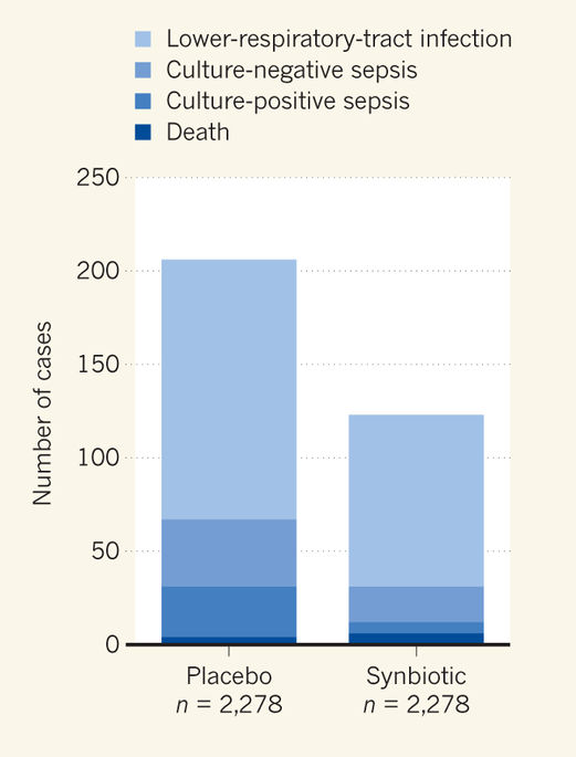
In this new publication, there were 72 babies who had MRI findings considered to predict a poorer outcome, and 22 with ultrasound findings that were considered predictive.

The study wasn’t designed this way but I would be very interested to see what happened to the families who had ultrasound findings not considered predictive, but MRI findings which were…

What the authors did actually do was to present the results of the un-masked test to the families.  This was done as follows “An experienced physician unaware of the unallocated result discussed the allocated images and neurodevelopmental prognosis in a structured interview with parents, providing permanent examples of the images and a written summary of the prognostic information”. The “structured interview” by an experienced member of this group, among the world’s experts in evaluation and interpretation of imaging findings in such patients, would have been very interesting to see more details about.

What should we say to parents when we have the results of a test with very poor positive predictive value for adverse outcomes, but relatively good negative predictive value? When the prevalence of that adverse outcome is relatively low? By which I mean that 92% of the infants in this study did not have CP with a GMFCS of 2 or more, similar to data in many other studies; if you have a normal pre-discharge MRI that increases to about 96%, (based on previous publications) a modest improvement in NPV over not doing an MRI, but still not to 100%.

I guess you would have to say something like “your baby has an imaging study which is normal/abnormal, which doesn’t have much impact on our prediction of their long-term outcome. He/She will be followed-up with frequent evaluations, and we expect that things will go well. There is a small chance of developing problems with control of movement, and your baby will be examined so that if something develops they can get any therapy that they need. There is about a 1 in 3 chance of a delay in language development, and a 1 in 5 chance of overall slower than average development, we will be doing testing to see if those things happen and can offer support and intervention if they do turn out to occur.”

I think you could say almost exactly the same thing to parents whose baby had an “abnormal” or “predictive” result on MRI or on ultrasound as you would say when the imaging does not show such abnormalities.

I don’t know what was said to the parents in this study, but they did not have an increase in their anxiety scores after the interview, indeed the scores went down a little in both groups. Anxiety scores were in fact the primary outcome variable of this study. The score used has a scale for “state”, that is how anxious you are feeling right now, and “trait”, that is how anxious you are in general over a period of time. Higher scores reveal more anxiety,  and various cutoffs have been suggested as indicating clinically important anxiety, such as over 39 for young adults to over 55 for geriatric patients, the minimum score possible is 20. A previous study of parents with a baby in the NICU showed trait scores averaging in the low 50’s.

In this study the mothers’ “state” anxiety scores were slightly higher in the ultrasound group before the imaging visit (by about 0.8 points), and stayed slightly higher (by about 1.5 points) throughout the follow-up to 18-24 months. A difference which was not likely due to chance (p=0.02). The “trait” anxiety scores were more similar between groups, and the fathers’ scores were not different between groups.

In other words, with a structured explanation of the results from an experienced individual who understands the limitations of the imaging tests, there is no adverse impact on anxiety. Both MRI and US were followed by a small decrease in anxiety, a decrease which was slightly greater in the MRI group.

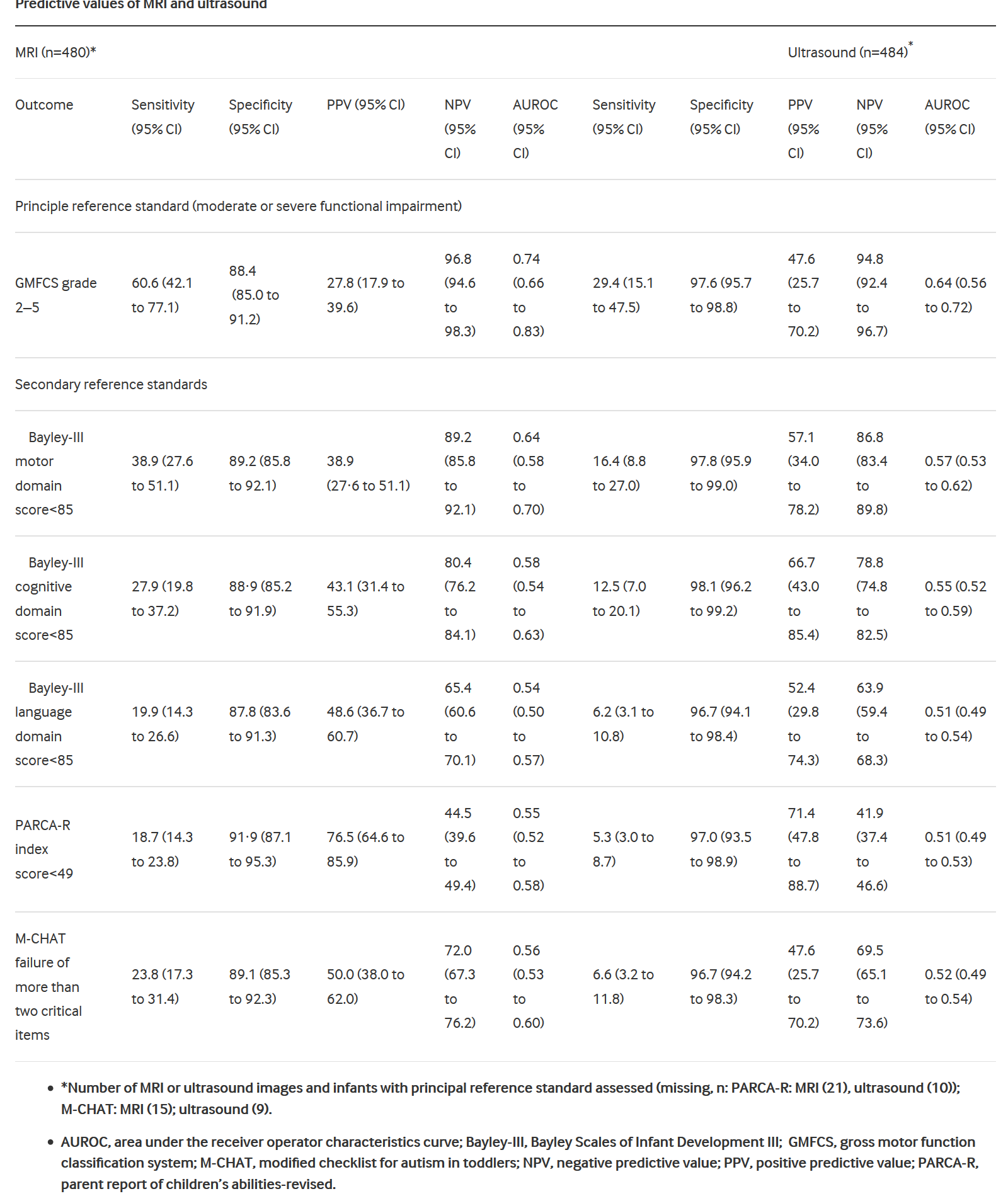
But were the tests actually useful? Were they actually predictive of long-term problems, based on Bayley version3 testing and neurological examination at 18 to 24 months corrected age? Here is table 4 from the publication showing the sensitivity, specificity and predictive values of MRI and ultrasound findings.

What they show basically is that the MRI finds more abnormalities, many of which are not associated with long-term problems, but some are. So the sensitivity of MRI is higher for all of the outcomes (but still relatively lousy, ranging from 18% to 60%) compared to head ultrasound (where they range from 5% to 16%), but the specificity is lower, around 90% for each outcome compared to around 97% for head ultrasound.

I think for an individual family the most important metrics are the predictive values, and, because of the increased details and increased detection of white matter abnormalities, the positive predictive values of an abnormal MRI are truly pathetic: from 28% to 48% for cerebral palsy and various components of the Bayley scores. The PARCAR-R test is a parent report of children’s abilities (revised), a parental questionnaire assessment which does not have the same standardization as the Bayley scales (i.e. it doesn’t have a mean of 100 and an SD of 15 in a standardized population), but a cut-off of 49 has been associated with a reasonably good distinction between infants with and without developmental delay, MRI had a very low sensitivity for this outcome, but was fairly specific, leading to a higher PPV but a low NPV. The M-CHAT is a screening test for autism features, which is affected by language delay; the MRI and the ultrasound prediction of failure on 2 or more items of the M-CHAT were quite similar to the results for prediction of language delay.

The authors of this article calculate the area under the  receiver operator characteristics curves, which were modestly higher for MRI than for US for prediction of moderate or severe motor dysfunction (0.74 compared to 0.64) and for motor dysfunction, but were very similar for cognitive and language dysfunction.

Finally they calculated costs, and the MRI cost about 300 pounds stirling more than the US.

I think this study confirms my prior evaluation of the usefulness of brain imaging at discharge of the very preterm baby. Sensitivity is poor for any modality of imaging. Specificity is higher for ultrasound, but is problematic for MRI.

Dr Edwards group and the developing human brain project (and other projects around the world) are incredibly important programs aimed at understanding how the human brain develops, and what can go wrong, so that eventually we will be able to figure out how to normalize brain development in more infants, term and preterm.

See these amazing images, for example, from the King’s College London human connectome project:

These are 3-D reconstructions of the cortical surface of newborn brains, with the images showing the surface of the white matter at the top, of the grey matter next, followed by the inflated surface (whatever that means) different structures, sulcal depth maps, brain curvature, cortical thickness and T1/T2 myelin maps. You can see the progression of complexity and maturation of the brain in the last 8 weeks of pregnancy.

Their detailed connectome images from developing brains are stunningly beautiful, showing the incredible profusion of connections within a developing brain.

Left: Multi-shell high angular resolution diffusion data decomposed into a free water component (greyscale background image) and a directionally resolved brain tissue component shown as rendered surfaces. Middle and right: Visualisation of anatomical connections in the developing brain derived from the brain tissue component.

MRI (or whatever you call these kinds of data maps, MRI seems much too simple a term) is an extremely important research tool, which is not the same as saying everyone should have an MRI!

Both term equivalent MRI and Ultrasound have very limited predictive ability for the former very preterm infant. This study shows, much more clearly than any before, that for an individual baby an MRI adds very little to an ultrasound, and that all former very preterm require health and developmental surveillance regardless of their imaging findings.

Posted in Neonatal Research | 4 Comments

Time to abandon the Papile classification? (part 3)

What should we do now?

I think we should stop using the Papile classification.

We should clarify that an intraventricular hemorrhage with acute hemorrhagic dilatation of the ventricle(s) is not the same pathophysiology as a hemorrhage followed by dilatation. (stage 2 followed by PHVD compared to a stage 3)

We should differentiate between an acute intra-parenchymal echodensity (which may be hemorrhagic or edematous) and PVHI, associated with intraventricular blood on the ispilateral side.

We should analyze and record the brain regions affected by the PVHI or IPE.

We should follow, and report the outcomes, of several hundreds of individuals, in order to have reliable information.

In other words, we should abandon the Papile classification, we should use descriptive terms to interpret head ultrasounds, we should use published percentiles to determine if a ventricle is dilated or not, we should record the brain regions affected on ultrasound, unilateral and bilateral, and we should work at correlating these findings with long-term outcomes.

Posted in Neonatal Research | 2 Comments

Time to abandon the Papile classification? (part 2)

As I mentioned in the last post, the initial report of outcomes by Bassan and others showed that the infants with severity score 0, (unilateral haemorrhage without midline shift affecting one zone) had reasonably good outcomes, 7 of 8 had cognitive scores within the normal range, 4 of 8 were without gross motor dysfunction. They also showed that having a VP shunt did not change significantly the neurological or developmental outcomes. As should therefore be obvious ‘grade 4 hemorrhages’ with these characteristics should be considered relatively benign, warranting close follow-up, and surveillance for complications such as ventricular dilatation or development of multiple cysts. Many PVHI resolve with a single porencephalic cyst in their wake, some resolve and leave findings similar to cystic PVL, that second group may well have worse motor outcomes than single porencephalic cysts, I will come back to this point later.

The Bassan study reports is obviously very small numbers, there were only 30 infants with PVHI in all (which isn’t bad for such a relatively uncommon lesion), so I asked myself if there are other similar data with larger numbers.

The largest I have found so far is a study from North Carolina (Maitre NL, et al. Neurodevelopmental Outcome of Infants With Unilateral or Bilateral Periventricular Hemorrhagic Infarction. Pediatrics. 2009;124(6):e1153-60) which has outcome data from 69 surviving infants with PVHI, 52 unilateral and 17 bilateral. The infants with bilateral PVHI were more immature than the unilateral babies, otherwise there was little difference in the early clinical course of the infants, but their outcomes at up to 36 months were very different.

Infants with unilateral PVHI had a Bayley II MDI at 18 to 36 months which averaged 82, compared to 49 with bilateral PVHI (almost all had an assigned score of 49). Cerebral palsy was much less common and much less severe with unilateral than bilateral PVHI, 15 of the 17 with bilateral PVHI had moderate or severe CP, compared to 19 of the 52 with unilateral PVHI. In concert with these findings the proportion with BayleyII scores above -2 SD on the MDI was significantly better with unilateral PVHI. In this study there is no description or analysis of the extent of the lesion on each side, or the worse side. They did report that those infants who had PVL on late ultrasounds had worse MDI, worse PDI, and more severe motor dysfunction than those who had porencephalic cysts.

Van Buuren et al (van Buuren LM, et al. Cognitive outcome in childhood after unilateral perinatal brain injury. Developmental Medicine & Child Neurology. 2013;55(10):934-40)
reported the outcomes of 21 babies who had unilateral PVHI, it is a somewhat unusual cohort in that the mean gestational age was 30 weeks, and ranged from 26 up to 41 weeks. One strength of the study is the later age IQ testing (performed, however, between 6 and 20 years of age) which showed that the children who had PVHI had full-scale IQ results which were lower than the standardizing population, but mostly within one standard deviation of that population, with a mean of 86. Verbal IQ scores were not significantly different from the theoretical norm at 96. Infants with post-hemorrhagic ventricular dilatation did worse, with a mean IQ of 80 compared to a mean of 96 for those without. The extent of the lesions is not clearly described they are noted as being frontal, fronto-parietal, or parietal and that the location had no impact on outcome, they don’t mention laterality (uni- vs bilateral); 12 of the 21 had a hemiplegia, but the severity is not described.

A study from Groningen (Roze E, et al. Functional Outcome at School Age of Preterm Infants With Periventricular Hemorrhagic Infarction. Pediatrics. 2009;123(6):1493-500)
followed 21 babies who had PVHI, there were 38 babies with this diagnosis in the cohort, 15 died and 2 were not included in the follow-up. They showed that only a minority had significant functional impairments, even though 9 of them had bilateral PVHI, and 5 had extensive (described as fronto-parietal-occipital) lesions. Although 16 children had Cerebral Palsy, 13 were GMFCS 1 or 2, only 3 had more severe motor problems. Most of the cognitive outcomes were normal or mildly affected with only about 10% more than 2 SD below the standardized mean at 4.4 to 12 years of age. They did not show an association between bilateral or more extensive PVHI and worse outcomes, but clearly the power is relatively low for these comparisons. Post-hemorrhagic ventricular dilatation was present in 8 infants, who had poorer scores, again the numbers are very small, but they seemed to have more cognitive difficulties.

The ELGAN cohort study included 54 babies with a diagnosis of PVHI. (O’Shea TM, et al. Neonatal cranial ultrasound lesions and developmental delays at 2 years of age among extremely low gestational age children. Pediatrics. 2008;122(3):e662-9). 44% of them had a Bayley II MDI less than 70 at 2 years of age, and 59% had a PDI under 70, there is no analysis of whether unilateral or bilateral PVHI were different, to unilateral or localised lesions.

A more complex system called the Abdi score is calculated as the square of the highest traditional IVH grade (so scoring 1, 4, 9 or 16), plus the IVH grade on the contralateral side, plus 5 for each hemisphere when more than two of its territories are involved, and plus 5 when there is a midline shift of the brain. The “territories” refers to the brain regions from the Bassan system. The score was first published in the Saudi Medical Journal and I can’t access the paper to see how it was derived, but as far as I can tell it was rather arbitrary. A recent outcome study using the Abdi scores (Al-Mouqdad M, et al. A New IVH Scoring System Based on Laterality Enhances Prediction of Neurodevelopmental Outcomes at 3 Years Age in Premature Infants. American journal of perinatology. 2016(EFirst) followed 183 very preterm babies at 3 years of age. They included 55 babies with intraparenchymal bleeds, whose Abdi scores could range from 16 (i.e. 4-squared) to 35 (4-squared plus 4 plus 5 plus 5 plus 5). The authors showed that  this score was better at predicting outcomes than simply using the Papile grade, but they did not compare to the Bassan system for PVHI. Although this score seems better for an individual patient prediction, it would be difficult I think to use it in daily practice, and the small numbers with each individual score mean that a very large database would be required to validate differences between each score

You may be asking yourself why is Keith Barrington/neonatalresearch.org going over all this older data? I was stimulated to do so by recent cases in my NICU and also by this publication from Texas (Sheehan JW, et al. Severe intraventricular hemorrhage and withdrawal of support in preterm infants. J Perinatol. 2017;37(4):441-7). The authors note that the long-term prognosis of very preterm infants with parenchymal hemorrhage has been shown to be much better than previously thought.

As you can hopefully see from this (non-exhaustive) review, the majority of infants with parenchymal bleeds (“grade 4 IVH” or PVHI) have outcomes which are within the range of healthy full term infants, only a minority have serious impairments (although, as most of the follow-up is only to 2 years of age, permanent functional impairments are impossible to quantify).  As you might expect, but with little data to support it, it seems likely that more extensive hemorrhagic lesions, and bilateral lesions have worse outcomes than limited or unilateral lesions, but there are very few patients who are informative in the published cohorts.

Sheehan’s study examined discussions and decisions regarding redirection of care among very preterm infants with a diagnosis on ultrasound of serious brain injury (“grade 3 IVH” or PVHI). They showed that the improved knowledge about the benign outcome of many of these lesions has not led to fewer decisions for comfort care. They also examined the outcomes of survivors with PVHI: 13/35 or 37% had no or mild NDI, 10 or 29% had moderate-to-severe impairment and 12 or 34% had profound impairment. Survivors with profound impairment had a median of 4 (IQR 3, 4) territories vs 2 (IQR 2, 3) territories in survivors without profound impairment.

Their data are therefore quite consistent with others, that parenchymal injury is often compatible with little or no impairment, and that the more severe the parenchymal injury the more severe the disability (although with wide confidence intervals). But this has not led necessarily to changes in decision making.

Why is this? In any other area of medicine, I suggest, if outcomes were shown to be much better (or at least, much less bad) than previous beliefs about long-term outcomes, decision-making would surely have changed, no?

I think we should reconsider our outcome data, and how good we are at predicting profoundly adverse outcomes.

What I suggest is the follows:

  1. The construction of robust large databases which relate head ultrasound findings to long-term, functional, outcomes. With enough detail to be able to relate a particular constellation of findings to a range of probable outcomes. Such databases will need to take into account mortality, but that must be predicated on whether care was re-directed or not. Withdrawal of life-sustaining interventions as a result of head ultrasound (or other considerations of long-term outcomes) must be factored into such databases.
  2. Development of tools to teach neonatal and perinatal trainees (and established caregivers) how to counsel parents regarding the results which would be routinely available from 1.

We must move on from a discussion focussed on “your baby has a grade 4 hemorrhage” and “life therefore is no longer worth living” (often implying, or overtly saying, that the long-term outcome will be “terrible”). To “your baby has these findings on imaging” which means that she (or he) will most likely be within the following range of outcomes, the majority of which are associated with an entirely acceptable quality of life.


Posted in Neonatal Research | Tagged , , , | 2 Comments