There are a huge number of publications correlating medium term outcomes (by which I mean outcomes around 1 to 2 years of age) with findings in the neonatal period. Most have concerned various approaches to brain imaging, although other studies have evaluated EEG, NIRS, early structured physical examinations, counting how many complications the baby had, the type of feeding they received…. I am sure my readers could construct a longer list.
There are several recent publications that have triggered this post, in the extremely preterm infant.
In the preterm infant, brain injury on imaging is very common, yet most preterm babies actually function very well. Why, therefore have we spent so much effort trying to refine brain imaging, to find better ways to predict outcomes?
The main point I want to make is the following:
Finding a statistically significant correlation is not the same as an individually useful prediction. Investigations seeking answers about the cause of neurological and developmental problems in the newborn might be satisfied to find a statistically significant correlation, that could be a reasonable research goal. But, just because there is a significant correlation doesn’t mean that a finding is useful to predict an individual child’s prognosis.
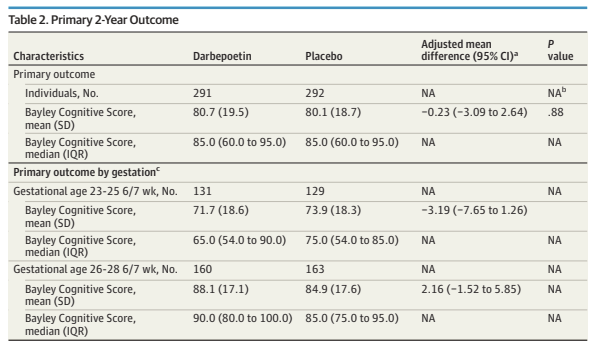
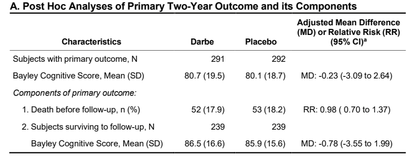
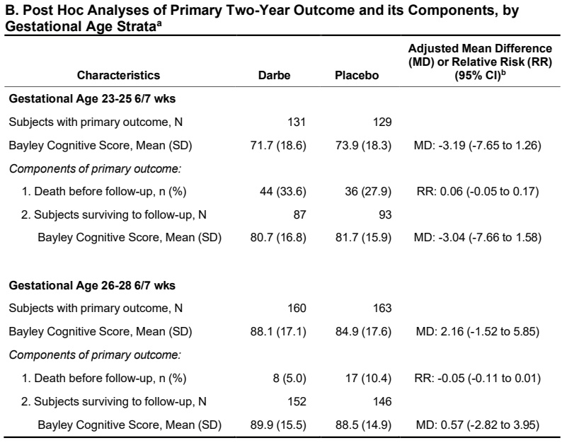
Let me give one older, illustrative, example, Tusor N, et al. Punctate White Matter Lesions Associated With Altered Brain Development And Adverse Motor Outcome In Preterm Infants. Sci Rep. 2017;7(1):13250. This study quantified the punctate white matter lesions (PWML) on MRI at term equivalent age, in a multicentre cohort of 500 preterm infants of <33 weeks. They examined the infants at 20 months corrected age, performed Bayley version 3 developmental screening and a neurological exam. 114 infants had PWML and a neuro exam, of whom 10 had CP with a GMFCS grade 2-5; 281 infants had no PWML and a neuro exam, of whom 2 had CP of those grades.
The incidence of CP among all those who showed up for a neurological examination, therefore, was 3.5%. If they had PWML on the MRI at term the incidence was 9%. So the difference in CP incidence was statistically significant, with an Odds Ratio of 6.6 (95% CI did not include 1, 2-22). But for an individual baby the finding was completely useless as a prediction!
If you found PWML on the MRI you could say with over 90% confidence to the parents, that the infant would not have disabling CP!
Even among those with PWML in the cerebro-spinal tracts, or those with large numbers of lesions, the individual prediction of CP was always under 30%.
Some years ago, an abnormal finding of what was called DEHSI, an abnormal white matter appearance of Diffuse Excessive High Signal Intensity, was reported as a frequent occurrence at term equivalent age in very preterm infants (Maalouf EF, et al. Magnetic resonance imaging of the brain in a cohort of extremely preterm infants. J Pediatr. 1999;135(3):351–7) it was initially thought to be a poor prognostic factor. However, further study suggested that it was not strongly associated with worse developmental outcomes. This new study, from a multicentre cohort of about 340 babies from Cincinnati of 32 weeks GA or less, (Derbie AY, et al. Diffuse white matter abnormality is independently predictive of neurodevelopmental outcomes in preterm infants. Arch Dis Child Fetal Neonatal Ed. 2025) used automated objective measurement of the signal intensity in the Centrum Semiovale. If you remember from your neuroanatomy, this is a large region of subcortical white matter superior to the lateral ventricles and the corpus callosum, which on each side of the brain is roughly the shape of half an oval! Increased signal intensity on T2 imaging, calculated by their algorithm as being more than 1.8SD higher than the mean density, they called Diffuse White Matter Abnormality DWMA. You can see from the outlined regions in the MRI below, you wouldn’t have spotted those regions by eye, it really requires their computerised algorithm.

These babies were then followed, with a good percentage returning for evaluation (nearly 90%), at 2 years for Bayley’s and neuro examination; at 3 years (also around 90%) they had cognitive testing done using a tool that is well validated, and somewhat predictive of later academic performance “the Differential Ability Scales, second edition (DAS-II) General Conceptual Ability (GCA) score”.
There was a strong correlation between several different factors, including the MRI findings, and motor scores, diagnosis of CP, and cognitive scores.
This kind of study always leaves me ambivalent. On the one hand, the finding that the extent of white matter injury correlates with motor, and, less strongly, with cognitive outcomes, is an unsurprising confirmation of the importance of abnormal brain development in very preterm infants. This is a very well done study, with infants from a wide range of gestational ages, excellent high quality follow up, and extensive statistical analysis.
On the other hand, what on earth are we supposed to do about it? The outcomes measured are largely outcomes that have little importance for parents. As the parents voices project has confirmed, they don’t care about Bayley scores, and CP with a GMFCS=3 is not considered by parents to be a serious adverse outcome.
In addition, the correlation with the outcomes is not even very close. The following figure shows the contribution of various factors to the cognitive outcomes of the infants. It shows that having antenatal corticosteroids (ACS), maternal breast milk (MMDD) and being a girl (SEX) had a substantial positive impact, whereas High-Risk Social Status (HRSS) had a major adverse impact, especially if combined with moderate or serious Brain Abnormalities (shown on the figure as msBA, defined by a Kidokoro score of over 7 on the term-equivalent MRI).

Those other factors were much more strongly correlated with the outcomes, than the DWMA. For example the relative impact of receiving antenatal steroids gave a ß value of +13.5 compared to about -2 for the DWMA, receiving maternal milk at discharge had a ß of +8 (which I think means that, after correcting for other factors, the cognitive score was on average 8 points higher among those who received maternal milk compared to those that did not, this is huge impact from something that we can do something about! Or, depending on how the statistics were done, it might be 0.8 SD higher: 1 SD was 20 points on the cognitive score, so 0.8 SD would be 16 points higher) As you can see from that graph above, the DWMA had a relatively weak association with cognitive score (all the results were similar for the motor composite score). The ß of about 2 means that, for each 1 SD increase in DWMA volume, there was an average of a 0.2 SD lower score on the Cognitive Composite, or 4 points lower.
Unlike some other markers on TEA brain imaging, the absence of DWMA does not appear to be very predictive of absence of CP, but I can’t find enough data to calculate the Negative Predictive Value in this publication.
I also find the way the results are presented to be questionable. The subtitles of the various sections of the results all use the term “prediction” such as in “prediction of motor performance”, “prediction of cognitive performance”, “prediction of CP”. These all presuppose that the prediction is useful, whereas they all were, in reality, very poorly predictive. It would have been better to title the sections “correlation with…”
There is a really good paper describing the methodology, goals, and interpretation of prognostic studies that I have been reading (Kent P, et al. A conceptual framework for prognostic research. BMC Med Res Methodol. 2020;20(1):172). It makes the important distinction between prognostic determinants and prognostic markers, and between various stages of exploratory and confirmatory research projects. This project would therefore be an exploratory study. Although the authors of this study have shown that there is a relatively weak association between the extent of DWMA and lower scores on the developmental screening tests at 2 to 3 years, we can’t tell from this whether the DWMA is a marker or is causative, and whether interventions aimed at reducing DWMA will improve outcomes.
To come back to the sentence in bold type above, the authors have shown a statistically significant correlation between having more white matter injury, using their algorithm on TEA brain MRI, and poorer motor scores at 2 years, lower cognitive scores at 3 years, and a diagnosis of CP (stage 1 or more). But, is that association useful in prognosis for an individual? They have shown that other factors are much more strongly associated with those outcomes, some of which are potentially modifiable on a group basis (working harder to ensure that mothers get steroids before delivery) or on an individual basis (maternal milk intake).
Another very recent study, from the same group, (Mahabee-Gittens EM, et al. Severity of punctate white matter lesions in preterm infants: antecedents and cerebral palsy prediction. Pediatr Res. 2025) analysed PWML, (the punctate lesions discussed above) and divided the 28 who had PWML into terciles of extent of lesions. 39 of the 339 infants in the study (12%) developed CP, of whom 6 were among those who had PWML.
To put it another way, the large majority of the babies who developed CP (33 of 39) did not have PWML. The majority of those with PWML, 22 of 28, did not develop CP.
This study also shows no significant correlation between the PWML and Bayley scores, on any of the 3 domains, including the motor composite.
This is a very unimpressive predictive capacity for the individual baby. Nevertheless, an accompanying editorial somehow uses those results to push the idea that all ex-preterm babies should have MRIs. They claim that doing so would allow early intervention, not mentioning that targeting early intervention to those with PWML on the MRI would fail to include the large majority of babies with CP. In fact, as usual, the study showed that the strongest predictor of poor developmental scores was Social Status, followed, in this analysis, by chorioamnionitis.
A much better way of targeting early intervention then, according to these data, is not to perform routine MRI, but to forget the MRI and routinely enrol infants with poor social status into early intervention programs.
Here’s another idea. Why not take the money that would be spent on routine MRI, and just give it to the poorest parents? (Bouchelle ZM, et al. Unconditional cash transfers to low-income preterm infants and their families: a pilot randomized controlled trial. J Perinatol. 2025) This pilot trial is investigating the impacts of giving unconditional money transfers to families that are at highest risk of having a baby with developmental delay. That is, according to the MRI study above, those with poor social status. Previous studies of giving money to families, with babies mostly born at term, (Gennetian LA, et al. Effects of a monthly unconditional cash transfer starting at birth on family investments among US families with low income. Nat Hum Behav. 2024;8(8):1514–29) showed that “mothers spent more time engaged in cognitively stimulating activities with their children. In addition, ~25% of the value of the cash gift was used on children’s books, toys, activities, clothing, diapers, and children’s electronic items/devices” which are all things likely to improve infant development, and which poor families struggle to obtain.
For 100 babies eligible for a TEA MRI, if they cost, say, $500 each, that $50,000 could instead be divided among the 10 families at highest social risk and the impacts on infant and family well-being might be dramatic. I think it would be ethically justifiable to have some minor conditions attached, such as participation in an early intervention program, which have been shown to improve cognitive and motor function in infancy (see the latest Cochrane Review Orton J, et al. Early developmental intervention programmes provided post hospital discharge to prevent motor and cognitive impairment in preterm infants. Cochrane Database Syst Rev. 2024;2(2):CD005495).
Instead of performing routine MRIs and searching for abnormalities with a weak association with CP or developmental delay, we should focus on ways to improve those delays and improve outcomes. The individual predictive ability of any finding on MRI at term-equivalent age is between low and extremely low. Such studies may well have value for research, but for improving outcomes of former very preterm babies they are useless.