When to deliver twins?

A new RCT from Australia randomized 235 mothers carrying twins to be either delivered at 37 weeks gestation, or allowed to continue for at least another week (Dodd J, Crowther C, Haslam R, Robinson J, for the Twins Timing of Birth Trial G: Elective birth at 37 weeks of gestation versus standard care for women with an uncomplicated twin pregnancy at term: The twins timing of birth randomised trial. BJOG 2012, 119(8):964-974 http://onlinelibrary.wiley.com/doi/10.1111/j.1471-0528.2012.03356.x/abstract).

The primary outcome variable was a composite of fetal/neonatal death or one of a number of serious morbidities, including asphyxia, immature lung disease, and fetal growth restriction ❤rd percentile. This is probably reasonable in general terms, but as with most composite outcomes the components are not of equivalent clinical importance, and clearly in this case are not mutually exclusive. A birth weight ❤rd centile is not of the same significance as asphyxia with encephalopathy.

Also, despite being a difficult trial that will most likely not be repeated, the power was not adequate to address the issue of respiratory distress and pulmonary hypertension as a result of cesarean section without labour at 37 weeks. They did not achieve the planned sample size, of about double the actual size, because of “lack of ongoing funding”. I can’t even tell from the article how many actually had a c/s without labour although half of the early delivery group (86% actually delivered between 37 weeks and 37 + 4 days, the rest before 38 weeks) had a labour induction, presumably the majority of the others had an elective section. As the serious respiratory complications are relatively uncommon, the study would have had to be much bigger to examine this effect.

However they did show that, if you wait and deliver most women after 38 weeks, twins aren’t growing very well at this point, and more of them fall below the 3rd %le, increasing from 3% in the early group to 10% in the “standard care” group. No consequences of being SGA, such as hypoglycemia are reported.

(of note there are a few irritating errors in the manuscript, including: the authors refer to stage 3 and stage 4 encephalopathy, and give the reference to Sarnat and Sarnat’s paper, which only describes 3 stages of course; the graph showing the distribution of gestational ages at delivery shows well over 100% of the early delivery mothers delivered before 38 weeks!).

The overall conclusion I guess is that there isn’t much obvious advantage to waiting until after 38 weeks to deliver twins, if you do you will have more babies fall below the 3rd %le. We could really have done with a much bigger trial that could have better addressed the respiratory risks, and focused on the mothers who did not get an induction, as having a trial of labour markedly reduces the respiratory consequences of being a little early, even if you are then delivered by c/s., but I won’t hold me breath for that to happen. We need some further very large observational studies to tell us a little more about those risks.

Finally you should note the frequency of assisted reproduction among the pregnancies, about 18% were the result of “assisted conception” a far higher rate than assisted conception among singletons, which is about 4% in Australia. They still have some way to go to control this adverse consequence of ART (although their latest report shows that they have been very effective at reducing the multiple pregnancy rate to about 8%), it is much worse in the rest of North America outside of Quebec the frequency of twins and tripets after IVF is about 30%. Here in Quebec we have reduced twin rates after ART from about 30% which is where we were a couple of years ago to under 5%. This can be done with a combination of government funding and strict regulation.
Posted in Neonatal Research | Tagged , , | 2 Comments

What is heteroresistance?

Recent cases of coagulase negative staphylococci, that were very difficult to eradicate, have led to an assessment of whether the phenomenon of heteroresistance may play a part. To be honest, for me this is entirely new; I was not aware of this phenomenon until very recently.

The idea is that colonies of the organisms involved in an infection have subpopulations that have much less susceptibility to antibiotics, in this instance to vancomycin. In the lab the germ will test sensitive to the antibiotic, but in reality it will be very difficult to eradicate the bug. In fact, what is seen is that about 1 in 100,000 to one in 1 million of the germs are more resistant to the antibiotic, if you sub-culture the susceptible strain you will again find that the germ is sensitive, but there is again a subgroup of about 1 in 100,000 to 1 in 1 million organisms that are much more resistant. Now, I hate not understanding things (except theoretical physics that I have accepted that I will never understand (but according to Richard Feynman, that is just fine because no-one does*)) so if anyone can explain the molecular basis of this, please let me know what is happening.

Anyway… these bugs seem to be sensitive on standard lab testing, but when specific ‘other tests’ are performed, there is again a subgroup that are resistant. It may be the first stage in evolution of antibiotic resistance.

A recent publication from France reports a very worrying trend of increased prevalence of heteroresistant coagulase negative staph (often referred to as CoNS). (Rasigade JP, Raulin O, Picaud JC, Tellini C, Bes M, Grando J, Ben Said M, Claris O, Etienne J, Tigaud S et al: Methicillin-resistant staphylococcus capitis with reduced vancomycin susceptibility causes late-onset sepsis in intensive care neonates. PLoS One 2012, 7(2):e31548.
http://www.plosone.org/article/info%3Adoi%2F10.1371%2Fjournal.pone.0031548#pone.0031548-Ng1)  The two other publications from NICUs that I have seen, also report this as a feature of S capitis, but in other patient populations, by which I mean adults, the phenomenon can affect not just capitis, but other CoNS species, such as S epidermidis and S hominis. And indeed the phenomenon was first noted with S aureus.

Clinically what is evident is persistent bacteremia despite good vancomycin serum concentrations, and often despite removal of central lines (Van Der Zwet WC, Debets-Ossenkopp YJ, Reinders E, Kapi M, Savelkoul PHM, Van Elburg RM, Hiramatsu K, Vandenbroucke-Grauls CMJE: Nosocomial spread of a staphylococcus capitis strain with heteroresistance to vancomycin in a neonatal intensive care unit. Journal of Clinical Microbiology 2002, 40(7):2520-2525.
http://jcm.asm.org/content/40/7/2520.abstract).

What to do about this is not yet totally clear, the first thing to do is to recognize it; standard sensitivity techniques do not work, specialized approaches are required. Once identified, do we just push up the vancomycin levels or add (or change to) other antibiotics, agents such as linezolid, daptomycin and tigecycline may be indicated. None of which I know much about, but linezolid use in the newborn has been reported a few times; there is some data about kinetics and toxicity, and it has been successful in clearing persistently positive cultures in reported cases, with low toxicity. There are reported cases of neutropenia in older patients, this has not been the case in newborns and young children and there are now reports of more than a couple of hundred young children and newborns treated with Linezolid in whom there was surveillance for neutropenia.

Linezolid is sometimes referred to as being bacteriostatic, but in reality the distinction between ‘static and ‘cidal antibiotics is of little clinical significance, if any. It is generally accepted that bactericidal activity may be important for treating meningitis and endocarditis, for other infections there is no proven advantage. Indeed the distinction between bactericidal and bacteriostatic drugs is quite unclear, many bacteriostatic drugs do kill bacteria, and bactericidal drugs often do not kill 100% of the germs within 24 hours. (Pankey GA, Sabath LD: Clinical relevance of bacteriostatic versus bactericidal mechanisms of action in the treatment of gram-positive bacterial infections. Clinical Infectious Diseases 2004, 38(6):864-870.
http://cid.oxfordjournals.org/content/38/6/864.abstract).

The paper that I referred to above, from Van Der Zwet, reports clonal spread of a heteroresistant CoNS in their NICU over a 4 year period until it was finally recognized. The prolonged positive cultures that are seen in such cases are a real worry, infants who have prolonged inflammation as a result of delay in eradication of CoNS have worse long term developmental outcomes, possibly as a result of the effects of the inflammatory mediators over a long period on the brain.

The important factors for our babies appear to be:

1. do whatever you can to reduce the prevalence of nosocomial sepsis.

2. that includes removing lines and reducing the duration of broad spectrum coverage

3. Make everyone wash their hands (this should be considered MANDATORY, anyone not washing their hands before touching a baby should be fired)

4. do whatever you can to reduce the prevalence of nososcomial… (OK I know I can be annoying at times)

5. when you have CoNS, consider testing for heteroresistance

6. eliminate the more resistant organisms with something (probably with one of the new antibiotics, but with the certain understanding that one day those fancy, (I hesitate to say clever because there is no intelligence required, it is just evolution) staph will become resistant to the new one as well).

7. Make everyone wash their hands, CoNS appear in the blood stream of our tiny fragile babies because they were on the skin of a health care worker, and the health care worker touched something, a tube, a catheter or the abdominal skin of a baby, without washing their hands well enough.

8. Make everyone wash their hands.

Isn’t it weird that in the same post I can talk about quantum physics and about how much we need to wash our hands?

* ‘If you think you understand quantum theory, you don’t understand quantum theory’. A frequently re-quoted quote, but it is not certain that he ever said it…

Posted in Neonatal Research | Tagged , | 1 Comment

Trying harder

One interesting recent publication, that I think may describe a phenomenon in medicine that has not previously been described, is from the NICHD network. This publication noted that there is a great deal of variation in outcomes, specifically survival without major morbidity, among NICUs in the network (Smith PB, Ambalavanan N, Li L, Cotten CM, Laughon M, Walsh MC, Das A, Bell EF, Carlo WA, Stoll BJ et al: Approach to Infants Born at 22 to 24 Weeks’ Gestation: Relationship to Outcomes of More-Mature Infants. Pediatrics 2012.
http://pediatrics.aappublications.org/content/129/6/e1508.long). As others have previously noted, the variation between units is bigger than the effect size of almost any recent neonatal innovation, nitric oxide or surfactant for example.

What this publication did was to compare the outcomes of different NICUS for babies which are all treated actively in all the units, that is babies of 25 weeks gestation and higher. They noted the above mentioned differences. They then examined the ways in which more immature (less than 25 weeks) infants were treated. What they noted were major differences in performance of cesarean sections at 22, 23 or 24 weeks, provision of antenatal steroids and active resuscitation in the delivery room. What they showed was that centers which were more active at 22 to 24 weeks gestation, had better survival without morbidity among the more mature infants, than centers that were less active with the extremely immature babies.

There are a few possible explanations: it could be that centers that were more active attracted lower risk mothers and babies; it could be that, as the mother and fetus progress from 23 to 26 weeks gestation, an active center has a more positive and active attitude all along the spectrum; it could be that being more active with the most difficult babies makes you better at dealing with babies who have fewer acute problems; it could be that centers that are less active at 22 to 24 weeks are more likely to withdraw care from more mature babies (most of the difference was in death, not other complications). I think from looking at the data that the latter seems less likely, but as the mode of death was not reported we can’t be sure.

My own bias from doing this job for many years is that, as you try harder at 23 weeks, you get much better at looking after 25 week infants. That may not be true, but it is consistent with my experience, and I am not sure if it has ever been shown in other domains of medicine. It makes sense that if you are prepared to operate on the most complicated neonatal heat diseases you will have better results with the more straight forward ones, if you try hardest to salvage severe trauma victims, the more moderately injured ones will be easier to save.

Does anyone know if there are data in other fields that are consistent with this guess? My gut feeling is that such data exist, but I don’t know about them.

Posted in Neonatal Research | 1 Comment

EEGs for preterm infants: Predicting Long Term Outcomes part 3

In some parts of the world EEGs are frequently performed in very preterm infants in the first few days of life as a screening test, and occasionally used to redirect care if abnormalities are found. Is this justified?

Are there abnormalities which are visible shortly after birth which are good predictors of profoundly abnormal long term disabilities? Is there some other valuable reason for performing routine EEG in preterm infants? The literature here is fortunately not huge (at least fortunate for someone wanting to review it!) compared to some other issues.

If we search for inception cohort studies of EEG in the first week of life in defined groups  of at risk preterm infants, with good follow up rates and adequate description of outcomes, we find relatively few studies. (Now of course EEGs may be required for evaluation of certain clinical findings, particularly possible seizures, I am talking about routine EEG for prognostication here, I am also talking about traditional multi-channel EEG, there will be another post sometime about amplitude integrated EEG).

There are two main findings on EEG which have been evaluated, background patterns and sharp transients, particularly rolandic sharp waves.

Generally speaking, there does not seem to be much evidence that background patterns are very predictive of long term outcomes. Now the background EEG patterns of the preterm infants are discontinuous. Periods of quiescence are interspersed with bursts of activity. The duration of the periods of suppression can be quatified as the interburst interval, and several papers have proposed norms for the duration of the IBI and of the bursts; unfortunately the norms proposed vary widely from one publication to the next, for example at 25 weeks an IBI of more than 30 seconds, 45, 60 and 90 seconds have all been proposed as abnormal.

One relatively recent study by Le Bihannic  (Le Bihannic A, Beauvais K, Busnel A, de Barace C, Furby A: Prognostic value of EEG in very premature newborns. Archives of Disease in Childhood – Fetal and Neonatal Edition 2012, 97(2):F106-F109 http://fn.bmj.com/content/97/2/F106.abstract) exemplifies some of the problems with this literature, they report EEG findings and outcomes of 61 infants of between 25 and 29 weeks gestation, 9 surviving infants from the same time period were not included. The follow up was of various durations and standardized testing was only done if the neurologist’s initial assessment found a problem (which I think is fine if you are only worried about serious long term consequences), there is no clear definition of severe or moderate impairment, except that the severely impaired children did not go to normal school, and moderate impairment included attention deficit, hyperactivity, and dyspraxia. The frequency of moderate and severe sequelae was well over 50%; 36, compared with 25 who were ‘normal’. The reported findings were that a dysmature EEG at some point during the hospitalisation had a reasonably good Positive Predictive Value for the combined outcome of severe or moderate sequelae. Now when moderately severe sequelae includes clumsiness (dyspraxia) I am not sure of the usefulness of this for clinical prediction, especially as the abnormalities on EEG were often only evident just prior to discharge at term equivalent age.

Another study from about 6 years ago, although with a relatively small sample of 32 preterm infants, also throws doubt onto the usefulness of term equivalent age EEG, (Randò T, Ricci D, Luciano R, Frisone M, Baranello G, Tonelli T, Pane M, Romagnoli C, Tortorolo G, Mercuri E et al: Prognostic value of EEG performed at term age in preterm infants. Child’s Nervous System 2006, 22(3):263-269.http://www.springerlink.com/content/l4914u8506086276)  background patterns at term were only weakly associated with long term problems. This paper notes some of the difficulties in interpreting the literature with previous studies describing with delayed maturation in the preterm EEG compared to term infants, equivalent maturation, or advanced maturation. 

Let us consider sharp transients on the other hand: temporal sharp transients are frequently seen in infants with all sort of major and minor problems including parenchymal injuries seen on ultrasound, they are only important it seems if they persist after the first week or so, and are numerous and high voltage. There is also some evidence that positive rolandic sharp waves, especially if of high voltage and frequent, are associated periventricular leukomlacia. With several studies suggesting they are very specific for PVL’, but the diagnosis of PVL is not always clearly defined, often persistent periventricular echodensity is included, which has a very poor inter-rater reliability. They are also said to be associated with the later development of abnormal motor function. But abnormal motor function in the largest series, from Marret and colleagues was a prospective cohort of 417 infants less than 33 weeks gestation who survived to be seen at 1 year (Marret S, Parain D, Menard JF, Blanc T, Devaux AM, Ensel P, et al. Prognostic value of neonatal electroencephalography in premature newborns less than 33 weeks of gestational age. Electroencephalography and Clinical Neurophysiology. 1997;102(3):178-85 http://www.sciencedirect.com/science/article/pii/S0013469496956556). Abnormal was described as either mild distal hypertonia or diplegia or tetraplegia at 12 months of age, and was seen in 108 infants. This is an extremely high incidence of PVL and of movement disorders in a relatively low risk population, making any external relevance questionable. Certainly the severity of some of these diagnoses must be questioned. There is also no evidence of a link between the rolandic sharp waves and cognitive outcomes.

Finally both PVL by these definitions and the rolandic sharp waves seem to be becoming less frequent, the paper by Le Bihannic noted above only found 2 EEGs with the finding.

One thing I haven’t discussed is seizures. Seizures are found occasionally in infants in these studies, and are usually associated with a poorer prognosis. Seizures are difficult to diagnose clinically in the preterm (and in the term infant) (Malone A, Ryan CA, Fitzgerald A, Burgoyne L, Connolly S, Boylan GB. Interobserver agreement in neonatal seizure identification. Epilepsia. 2009;50(9):2097-101. http://onlinelibrary.wiley.com/doi/10.1111/j.1528-1167.2009.02132.x/abstract.) and are often under-diagnosed and mis-diagnosed.  More accurate diagnosis should lead to more appropriate treatment, and, one would hope, better outcomes (although that is not certain). So studies focused on seizure diagnosis and therapy have a potential to actually improve, rather than just attempt to predict, clinical outcomes.

So overall I don’t think there is enough evidence of accurate prediction of outcomes to warrant routine EEG monitoring of the preterm infant for that purpose, rolandic sharp waves if frequent (I didn’t go into it but there are 2 types and only type 1 seem important) may be highly predictive of PVL, but a confirmation of PVL with imaging would be required anyway, and they don’t correlate well with severity of outcome.

Posted in Neonatal Research | Tagged , | Leave a comment

Pulmonary compliance changes after surfactant

This blog is a bit different: not a review of a recent publication, but rather a teaching point for my junior trainees (and maybe some seniors).

The question is this: when you have preterm infant with typical HMD and respiratory distress in the first few hours of life to whom you give surfactant; what happens to pulmonary compliance? Try not to look at the answer until your knee has jerked and you have made your choice.

The correct answer is not it increases but ‘it depends’ (that is often the correct answer) but on what?

If you answered it depends on whether you are referring to static or dynamic compliance, then you clearly have either some insight into pulmonary physiology, or you made a lucky guess. Let me explain a bit more:

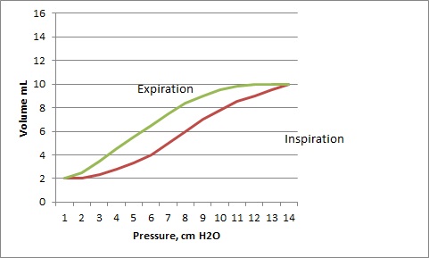
Static compliance is measured when there is no gas flow (hence static) and to produce the complete pressure volume loop the applied pressure should be taken down to zero. This gives the typical curve with its hysteresis.

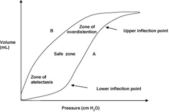
When you look at the display on a modern neonatal ventilator what you see is nothing like the curve shown above, what you see looks more like a dynamic pressure-volume curve (of course it does, that is what it is!)

Dynamic compliance is measured during tidal breathing (or tidal ventilation) so delays in gas movement as a result of airway (or endotracheal tube) resistance are important, and the curves look quite different.

The dynamic Pressure-Volume Curve looks like this

  • The usual effect of giving surfactant is an improvement of STATIC lung compliance. (Stenson BJ, Glover RM, Parry GJ, Wilkie RA, Laing IA, Tarnow-Mordi WO: Static respiratory compliance in the newborn. III: Early changes after exogenous surfactant treatment. Arch Dis Child Fetal Neonatal Ed 1994, 70(1):F19-24. http://fn.bmj.com/content/70/1/F19.long)
  • Now if static compliance is improved: and you make no changes on the ventilator, that is you have the same PEEP, then end-expiratory lung volume is higher.
  • This shifts the lung up the pressure-volume curve, and the end-inspiratory portion is likely to be on a flatter part of the curve

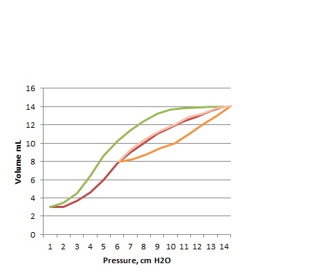
A before and after look at Static compliance loops during surfactant treatment look like this:

BEFORE, An applied pressure increasing from 0 to 13 cmH2O gives a volume of 8 mL.

CLstat = 8mL/13 cm H2O before surfactant: 0.66 mL/cmH2O

AFTER, An applied pressure increasing from 0 to 13 cmH2O gives a volume of 11 mL

CLstat = 11mL/13 cm H2O after surfactant: 0.85 mL/cmH20

But during tidal ventilation what we see and measure are DYNAMIC loops, such as these before and after loops below.

Pre-surfactant

And Post-Surfactant

If you look at the pressure volume loops on the front of your new fancy ventilator, that cost an extra 10,000 dollars for you to have pressure volume loops displayed, they will look like this, that is, there is no difference between pre and post surfactant loops. What you will also always see is that the end-expiratory lung volume is zero. Neonatal ventilator graphics reset to zero at the end of each breath, this has to be so, because of ETT leaks, if the graphics did not reset they would soon disappear off the screen.

So now we see that the Dynamic compliance CLdyn is 6 mL/8 cmH2O both before and after surfactant.

If we superimpose these dynamic loops on the same static curves that I constructed above you can see what has happened:

The first is Pre-surfactant:

The next is post surfactant:

The end-expiratory lung volume is higher, the tidal ventilation is now occurring at a higher part of the Static pressure volume relationship, and therefore at a flatter part. So if you measure dynamic compliance shortly after giving surfactant and before making any ventilator changes you see no improvement, this has been demonstrated multiple times. Here is one example of dynamic compliance before and 1 hour after surfactant therapy. (Couser R, Ferrara T, Ebert J, al. e: Effects of exogenous surfactant therapy on dynamic compliance during mechanical breathing in preterm infants with hyaline membrane disease. The Journal of Pediatrics 1990, 116(1):119-124. http://www.sciencedirect.com/science/article/pii/S0022347605816609)

  Surfactant Control
  Before After Before After
Compliance (ml/cm H20/kg) mean (SD) 0.43 (0.21) 0.45 (0.45) 0.33 (0.14) 0.33 (0.13)

This means that you cannot use the pulmonary graphics to determine whether the PEEP is optimal or if the baby is ready to wean. A better way to determine whether the surfactant has had an effect is simply to watch the FiO2; as the figure in Ben Stenson’s article referenced above shows, the decrease in FiO2 occurs at the same time as the improvement in Static Compliance. So when the FiO2 falls, reduce the PEEP: you will immediately afterward see the pip fall (on volume ventilation) or the volume increase (on pressure ventilation), and now you will be able to note the improvement in compliance.

Infants who reduce to 21% after surfactant can be managed with a reduction in PEEP to as low as 3 cmH2O (Dimitriou G, Greenough A, Laubscher B: Appropriate positive end expiratory pressure level in surfactant-treated preterm infants. Eur J Pediatr 1999, 158:888-891 http://www.springerlink.com/content/jumq39g0c58auql7/?MUD=MP) or even to 2 cmH2O (Sandberg KL, Silberberg AR
What Is the Best Positive End Expiratory Pressure (PEEP) in Ventilated Low Birth Weight Infants? PAS-meeting abstracts 2009, #3858.115).

Please don’t tell me that this is ‘below physiologic PEEP’ as many residents have told me in the past. There is no such thing as physiologic PEEP. Positive pressure ventilation is not physiologic. The physiologic pressure in the airways at the end of expiration is zero. If it were not so, it would not be the end of expiration.

So the actual optimal PEEP would be what allows airways and airspaces to stay patent, and at the same time avoids overdistension. It is unlikely that there is one pressure which will suit all babies, but PEEP which is very low can be optimal for some babies, you can tell if you have gone too low when the FiO2 starts to increase again.

So when you give surfactant, don’t expect a change in the pressure volume loops, expect the FiO2 to decrease, when this occurs decrease the PEEP, and if the baby stays in 21% you can decrease to 3 cmH2O. Then get the baby extubated!


Posted in Neonatal Research | Tagged , , | 3 Comments

This is what it’s all about: Bad Astronomy

This has nothing directly to do with neonatology. I just thought it was a wonderful 5 minute explanation of what science is all about. Thanks to Phil Plait at his blog Bad Astronomy. It is a brief speech he made at a Science Fair.

http://blogs.discovermagazine.com/badastronomy/2005/05/09/science-fare/

I know a place where the Sun never sets.

It’s a mountain, and it’s on the Moon. It sticks up so high that even as the Moon spins, it’s in perpetual daylight. Radiation from the Sun pours down on there, day and night, 24 hours a day — well, the Moon’s day is actually about 4 weeks long, so the sunlight pours down there 708 hours a day.

I know a place where the Sun never shines. It’s at the bottom of the ocean. A crack in the crust there exudes nasty chemicals and heats the water to the boiling point. This would kill a human instantly, but there are creatures there, bacteria, that thrive. They eat the sulfur from the vent, and excrete sulfuric acid.

I know a place where the temperature is 15 million degrees, and the pressure would crush you to a microscopic dot. That place is the core of the Sun.

I know a place where the magnetic fields would rip you apart, atom by atom: the surface of a neutron star, a magnetar.

I know a place where life began billions of years ago. That place is here, the Earth.

I know these places because I’m a scientist.

Science is a way of finding things out. It’s a way of testing what’s real. It’s what Richard Feynman called “A way of not fooling ourselves.”

No astrologer ever predicted the existence of Uranus, Neptune, or Pluto. No modern astrologer had a clue about Sedna, a ball of ice half the size of Pluto that orbits even farther out. No astrologer predicted the more than 150 planets now known to orbit other suns.

But scientists did.

No psychic, despite their claims, has ever helped the police solve a crime. But forensic scientists have, all the time.

It wasn’t someone who practices homeopathy who found a cure for smallpox, or polio. Scientists did, medical scientists.

No creationist ever cracked the genetic code. Chemists did. Molecular biologists did.

They used physics. They used math. They used chemistry, biology, astronomy, engineering.

They used science.

These are all the things you discovered doing your projects. All the things that brought you here today.

Computers? Cell phones? Rockets to Saturn, probes to the ocean floor, PSP, gamecubes, gameboys, X-boxes? All by scientists.

Those places I talked about before? You can get to know them too. You can experience the wonder of seeing them for the first time, the thrill of discovery, the incredible, visceral feeling of doing something no one has ever done before, seen things no one has seen before, know something no one else has ever known.

No crystal balls, no tarot cards, no horoscopes. Just you, your brain, and your ability to think.

Welcome to science. You’re gonna like it here.

Posted in Neonatal Research | Leave a comment

Long Term outcomes after Therapeutic Hypothermia for HIE

Two new publications from multicenter trials of hypothermia as a treatment for post-asphyxial encephalopathy. The first from the NICHD network, and the whole body cooling study led by Seetha Shankaran. They examined the children and performed IQ testing at 6 to 7 years. (Shankaran S, Pappas A, McDonald SA, Vohr BR, Hintz SR, Yolton K, et al. Childhood Outcomes after Hypothermia for Neonatal Encephalopathy. New England Journal of Medicine. 2012;366(22):2085-92http://www.nejm.org/doi/full/10.1056/NEJMoa1112066#t=abstract) There were 208 infants originally in the trial and only 18 of the surviving 140 infants were lost to follow-up.

The results confirm that therapeutic hypothermia improves survival without disability, diagnosing disability if the IQ was less than 70 (although it was not quite ‘significant’; p value was 0.06) and a reduction in severe disability (which was IQ less than 55, disabling cerebral palsy or blindness).

The second was a follow-up of the CoolCap trial, the parents were telephoned in order to complete the WeeFIM, a functional classification of the children’s abilities, at 7 to 8 years of age. (Guillet R, Edwards AD, Thoresen M, Ferriero DM, Gluckman PD, Whitelaw A, et al. Seven- to eight-year follow-up of the CoolCap trial of head cooling for neonatal encephalopathy. Pediatr Res. 2012;71(2):205-9. http://www.nature.com/pr/journal/v71/n2/full/pr201130a.html) Unfortunately only half of the babies were followed, which reduces the reliability of the results. (As an aside Pediatric Research now puts the methods at the end of the abstract, and at the end of the paper in smaller type. I think this is just weird. The methods are the most important part of any paper, how can you make anything out of the results if you read them before the methods? Can we start a campaign to make them change this back?)

What is really strange also is that the authors do not present the results from the cooled versus the control groups. Although they appropriately note that they had limited power because of the large numbers of drop-outs, I don’t understand why they didn’t at least present the data in that way, they focus instead on the predictability of the WeeFIM from the 18 months Bayley scores. I don’t see that as being any less affected by the low follow-up rate, but the authors claim that the data show that the Bayley is a relatively good prediction of functional outcome at 7 to 8 years. In fact though when I look at their figure 3 it seems that there are quite a few infants with an 18 month Bayley of 70 or less who have quite good functional scores.

If we examine the same relationship from Shankaran’s paper (looking at the supplementary material in the on-line appendix table 3) we can see that of 36 infants identified with a low Bayley at 18 months, 33 of them had an IQ less than 70 at 6 to 7 years. And of 86 infants without low Bayleys then 76 had an IQ over 70.

This is a much higher positive predictive value of an 18month Bayley in this study (even though the numbers are becoming quite small) than is the case for the preterm infant. It could make some sense that the cerebral injury in asphyxia is more diffuse, leading to a lower capacity for the plasticity of the brain to overcome the injury, whereas in preterm infants the injuries may be more patchy, especially periventricular hemorrhage, so they are more likely to slowly improve and catch up.

This confirms the value of hypothermia,  which has now been studied in a large number of similar trials, recently an updated systematic review was published, (Tagin MA, Woolcott CG, Vincer MJ, Whyte RK, Stinson DA. Hypothermia for Neonatal Hypoxic Ischemic EncephalopathyAn Updated Systematic Review and Meta-analysisHypothermia for Neonatal Encephalopathy. Arch Pediatr Adolesc Med. 2012;166(6):558-66. http://archpedi.jamanetwork.com/article.aspx?doi=10.1001/archpediatrics.2011.1772 ) Tagin and colleagues included RCTs that had follow-up to at least 18 months of age, which meant 7 trials with a total of over 1200 infants. This analysis confirmed that hypothermia decreases death, and decreases neurologic abnormalities and low Bayley scores.

I have only one beef with this well done meta-analysis: the very weak recommendation at the end of the abstract: ‘Clinicians should consider offering therapeutic hypothermia as part of routine clinical care to these newborns’.

Should ‘CONSIDER’ ?!

How about ‘Clinicians who do not offer therapeutic hypothermia are negligent’! We should sometimes go beyond the limits of the usual niceties of standard scientific writing when we are talking about saving babies lives and reducing handicap. Fortunately I don’t have an editor here on my blog!

Posted in Neonatal Research | Tagged , , , | Leave a comment

Desperately Seeking Problems with Premies

I don’t know about you, but I am getting really irritated with people searching desperately to find problems with the long term outcomes of preterm infants.

A new publication exemplifies this. A linked database study from Sweden has compared the frequency of hospitalisation for serious psychiatric disorders according to the gestational age at birth. (Nosarti C, Reichenberg A, Murray RM, Cnattingius S, Lambe MP, Yin L, MacCabe J, Rifkin L, Hultman CM: Preterm Birth and Psychiatric Disorders in Young Adult LifePreterm Birth and Psychiatric Disorders. Archives of General Psychiatry 2012, 69(6):610-617.) http://archpsyc.jamanetwork.com/article.aspx?doi=10.1001/archgenpsychiatry.2011.1374

The first question to ask is whether the data appear likely to be reliable. The subjects were born between 1973 and 1985, at a time when few women were getting antenatal ultrasounds, so individual errors are likely to be frequent, but the overall distribution of gestational age should be OK. But, the distribution of gestational ages is highly suspect: 17% of the deliveries were at 42 weeks or more, whereas 4.1% were less than 37 weeks and 0.4% less than 32 weeks, this should immediately ring alarm bells about the reliability of these data. These distributions are so far from what we would expect to see; either there has been a huge change in human biology over the last 30 years, or these gestational ages are systematically wrong.

But if we were to assume for a moment that the gestational age data were reliable, what do they actually show?

Well let’s focus on what is supposed to be the most striking finding, an enormous increase in bipolar disorder, relative risk among infants less than 32 weeks of 7.2, ‘fully adjusted’ relative risk of 7.4.

If we examine these numbers a little closer, rather than looking at the ‘relative risk’ let us examine the actual frequencies. Among full term infants there were about 150 hospitalisations among just over 1 million individuals. Among infants of less than 32 weeks gestation, there were ….. wait for it… 4 hospitalisations among 5125 individuals. That’s right, this paper, considered worthy of a news item in the BMJ, this paper, which makes broad general comments about brain development in the preterm and how surveillance of preterm infants should continue for life, this paper is based on 4 cases of hospitalisation among over 5000 preterm patients.

In addition, apart from the unreliability of the gestational age data, the authors had little information to adjust the frequencies for other risk factors, many of which could easily differ between premature and full term individuals.

So even if it were all true, the actual attributable risk of being preterm is 1 admission for bipolar disorder per 10,000 patient years. Now as an exercise in epidemiology, perhaps there is some value here, if there is some tiny increase in risk of serious psychiatric disorder among preterm infants that could have some importance (though I have difficulty imagining what it is), but to state, as these authors do, that these data show some general abnormalities in brain development among preterm infants is ridiculous.

If we add together all of the disorders that the authors claim to be significantly related with prematurity, we find that the term individuals had 3,114 hospitalizations for depression, nonaffective psychosis, bipolar disorder and eating disorders, out of the same million people. The premature infants had 41 out of 5,125. Sure the relative risk is increased, but the actual attributable risk is about 10 admissions more for every 10,000 patient years.

Rather than the ludicrous generalizations that the authors make about premature infants brain development, and need for surveillance, these data (even if they were reliable, which I doubt) say exactly the opposite. as far as serious psychiatric illness is concerned there is almost no difference between premature infants and the full term.

Posted in Neonatal Research | Tagged | Leave a comment

Head Ultrasound lesions and developmental progress: Predicting Long-Term Outcomes part 2

Two recent articles that give much more information about the predictive capacity of head ultrasounds.

Many articles about head ultrasounds and outcomes use the Papile classification. This classification was developed for simple classification of periventricular hemorrhage types as seen on CT scan into broad categories. The fourth of of those broad categories is extremely broad.

There are 2 major problems with the current use of the Papile classification. The first being that a modestly sized intraventricular hemorrhage, which subsequently develops ventricular dilatation, may be referred to as a grade 3 hemorrhage. A large intraventricular hemorrhage which acutely dilates the ventricles is also referred to as a grade 3 hemorrhage. These 2 occurrences do not necessarily have the same consequences, and should be differentiated, but in many publications it is not clear whether such a discrimination has been made.

More importantly I think, a grade 4 hemorrhage can refer to a multitude of different injuries. A small localized intracerebral hemorrhage, and huge bilateral hemorrhagic destruction of the hemispheres are both referred to as grade 4 hemorrhage, and subsequently grouped together in outcome studies. It is self-evident that they must have different consequences for brain function, but finding the relative significance of these 2 variations is next to impossible in the literature.

It also seems very likely that frontal, parietal, temporal, and occipital hemorrhages may have different effects, but again there has been little published information to use to inform parents. Furthermore, an echodense lesion may appear to be a hemorrhage to one person, but may be thought to be infaction or oedema to another.

This huge problem has been addressed by the ELGAN study (a multi-center prospective cohort study of Extremely Low Gestational Age Newborns); that is, inter-rater variability in interpretation of head ultrasounds, both in how the findings are perceived, and how they are reported. By variations in how they are perceived I mean that even when sonologists are asked to just determine whether there is a parenchymal lesion, (which they can then note as being an echodensity or echolucency), they are often unable to agree. In one report from the study (Kuban K, Adler I, Allred E, Batton D, Bezinque S, Betz B, Cavenagh E, Durfee S, Ecklund K, Feinstein K et al: Observer variability assessing US scans of the preterm brain: the ELGAN study. Pediatr Radiol 2007, 37(12):1201-1208. http://www.springerlink.com/content/k2637421vj4vvq74/?MUD=MP) they noted that when one sonologist reported an echodense lesion, about 50% of the time the lesion was not reported by another sonologist. Similarly, about 40% of echolucent lesions were not reported by a second sonologist. We often make critical decisions based on these findings, so these discrepancies are very concerning.

Similarly variable are the terms used by sonologists to report the same lesions. The same lesion may be referred to as early PVL, late PVL, or periventricular hemorrhagic infarction, by different sonologists. (Westra S, Adler I, Batton D, Betz B, Bezinque S, Durfee S, Ecklund K, Feinstein K, Fordham L, Junewick J et al: Reader variability in the use of diagnostic terms to describe white matter lesions seen on cranial scans of severely premature infants: The ELGAN study. Journal of Clinical Ultrasound 2010, 38(8):409-419. http://onlinelibrary.wiley.com/doi/10.1002/jcu.20708/abstract)

For this reason, the ELGAN study decided to describe simply whether or not there was an echolucent or echodense lesion; but as noted above this did not eliminate the differences in interpretation.

To get back to the two recent publications referred to at the start of this post:

Michael O’Shea and the ELGAN study investigators (O’Shea TM, Kuban KC, Allred EN, Paneth N, Pagano M, Dammann O, Bostic L, Brooklier K, Butler S, Goldstein DJ et al: Neonatal cranial ultrasound lesions and developmental delays at 2 years of age among extremely low gestational age children. Pediatrics 2008, 122(3):e662-669. http://pediatrics.aappublications.org/content/122/3/e662.long) have compared the location of echodense and echolucent lesions and the neurodevelopmental outcomes at 2 years of age, according to neurologic exam and Bayley 2 MDI and PDI.

They have shown that lesions in different areas of the brain have different impacts on the likelihood of low Bayley MDI and PDI scores. One example is the figure shown below showing the effects of an echolucent lesion in different locations on the scores on a 2 year Bayley exam (the proportions with an MDI <70 are on the left, PDI <70 on the right:

Percentage of children whose scan had an echolucency in a particular location and who had an MDI of <70 (black numbers on the left side of the brain) or a PDI of <70 (black numbers on the right side of the brain) on the BSID-II

The other article from Stephanie Merhar and others (Merhar SL, Tabangin ME, Meinzen-Derr J, Schibler KR: Grade and laterality of intraventricular haemorrhage to predict 18-22 month neurodevelopmental outcomes in extremely low birthweight infants. Acta Paediatr 2012, 101(4):414-418. http://onlinelibrary.wiley.com/doi/10.1111/j.1651-2227.2011.02584.x/abstract ) has examined the association between whether periventricular hemorrhages were unilateral or bilateral and Bayley Scores. They found several interesting things. Most importantly infants with grade 4 hemorrhage (referred to as PVHI in the ELGAN studies) did NOT have worse Bayley scores at 2 years, unless the lesion was bilateral. Infants with unilateral grade 4 hemorrhages had a range of Bayley MDI scores which was very similar to those with unilateral subependymal hemorrhage.

Secondly, even among those with bilateral PHVI if they did not develop either sepsis or receive postnatal steroids, then only 25% had an MDI less than 70. In contrast, those who had Bilateral PVHI and had either sepsis or steroids or both had an increasing proportion of children with low Bayley scores.

To me this means that the preterm brain has a huge potential for repair and recovery. When a serious injury occurs, even if a large part of the brain is affected,  there is a chance of recovery and good outcome, but if that repair is interrupted by sepsis or dexamethasone, then the outcomes are much worse. These results should make us pause and reflect: bilateral parenchymal hemorrhages do not necessarily lead to an MDI less than 70, even those with an MDI less than 70 will often have IQ in the “normal” range in later life. Very few will be profoundly multiply disabled. The addition of multiple other insults during hospitalisation progressively worsens outcome.

Posted in Neonatal Research | Tagged , | Leave a comment

Predicting Long Term Outcomes, General Observations

Just back to blogging from a few days at a very successful conference in Geneva, organized by Annie Janvier. At the conference I gave a presentation about prediction of long term outcomes in preterm infants. Some of the issues that I raised will be the subjects of a series of posts over the next couple of weeks, so I will be discussing the usefulness of ultrasounds, MRIs, EEGs, amplitude integrated EEGs, clinical examinations and clinical complications and anything else that has been investigated as possibly being of value in predicting long term outcomes among preterm infants.

A few general observations to start with:

Most reports that you can analyze to determine the reliability of a particular test or finding are flawed, and flawed in one very important way. The usually do not report the number of  patients with a particular finding during their hospital course who either died, or had active therapy withdrawn or withheld before they reach follow up. So for example we may have information about the infants with intraparenchymal hemorrhage who reached 2 years of age, but we often do not know about those, who may be a substantial proportion, who acutely died or had palliative care. This could be important, if those who had care withdrawn or died were systematically different from those who had on-going care.

Indeed I think it is likely that there are such systematic differences. I think it is likely that in some centers many or even most infants with intraparenchymal hemorrhage were offered redirection of care. Meaning that the parents of those who refused will have had a different background or different motivations to the rest. I think it is likely that in other centers withdrawal of active care has been offered selectively, meaning that those with less severe lesions are more likely to have survived. I think it is likely that in other centers redirection of care is not the primary consideration of the neonatologists, and it is parents who make the suggestion, again the characteristics of the parents will differ between those who survive and those who do not.

You will note that I use a phrase 3 times in that paragraph that I rarely use in anything I publish “I think it is likely”, I try to be as evidence-based as possible, but in this instance I cannot be. I am not aware of data about differential characteristics of patients and families whose babies survive with significant brain injuries (or significantly abnormal EEG or anything else) and those who die. Or whether the brain injuries differ between survivors and non-survivors.

I think in the future that studies of early prediction of outcomes of preterm infants should report the total cohort, those who had the findings of interest, those who died, those who had care redirected and those who survive. They should report the findings on the particular test of each category of patient to demonstrate whether survivors really represent the group of all those who had that finding. If not we could be really misled by the data.

Posted in Neonatal Research | Tagged | Leave a comment