Should we ever close the PDA?

The latest large multicentre RCT has just been published. Laughon MM, et al. Expectant Management vs Medication for Patent Ductus Arteriosus in Preterm Infants. JAMA. 2025. In this trial, infants of 22 to 28 weeks GA were randomized at between 48 hours and 21 days of life after an echocardiogram. They were classified into: 1. no or asymptomatic PDA; 2.symptomatic PDA; or 3. cardiopulmonary compromise. Only group 2 were randomized. The definitions are shown below, including, at the bottom of the figure, the definition of group 2. Of note, infants receiving hydrocortisone were ineligible.
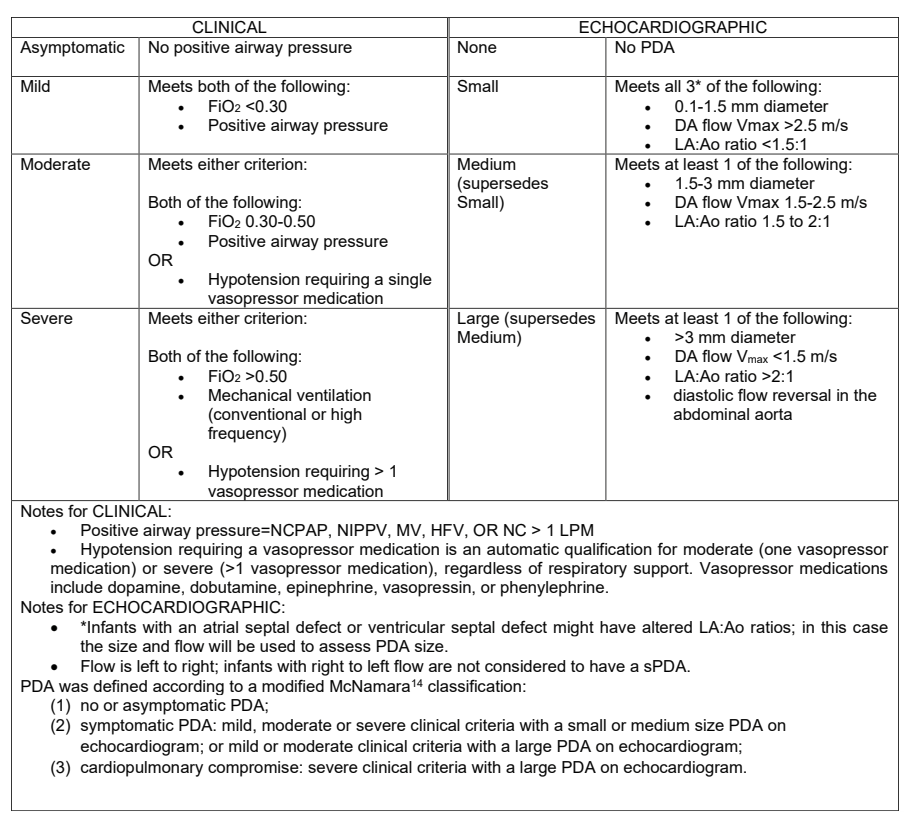
481 infants were randomized, within 48 hours of being eligible, and at a median of 10 days (IQR7-14) of age to either medical treatment, with ibuprofen, indomethacin or acetaminophen, or to control, expectant management. Controls were not supposed to receive any of those drugs unless they progressed to group 3 (cardiopulmonary compromise) or reached 36 weeks. There were unfortunately a large number of protocol violations, 60, or 25% of control, expectant management, infants received medical intervention (or surgical/catheter closure) for their PDA, of which 44 did not meet the agreed treatment criteria, and were therefore protocol violations.

I find this a little hard to understand, why get involved in the study if you are not prepared to abide by the protocol? Nearly 1 in 5 expectant group babies had PDA closure attempted even though they were not in the category of having severe clinical criteria with a large PDA, and therefore did not satisfy the protocol indications for treatment.

The primary outcome criterion was survival without BPD. I know, don’t get me started, designing a trial with a dichotomous outcome, that equates death with being in oxygen at 36 weeks, would be ridiculous in this day and age, in 2025, when so much better ways of designing trials with potentially conflicting outcomes, and analysing them in ways that take into account the relative importance of the outcomes, exist, and are now being used in other fields. It also leads to other rather, er, questionable decisions, such as defining death as death up to 36 weeks PMA. Really? Who cares about death up to 36 weeks, so an infant who dies at 37 weeks wasn’t counted in this trial?

The trial was, strictly speaking, a null trial. The primary outcome was identical between groups.
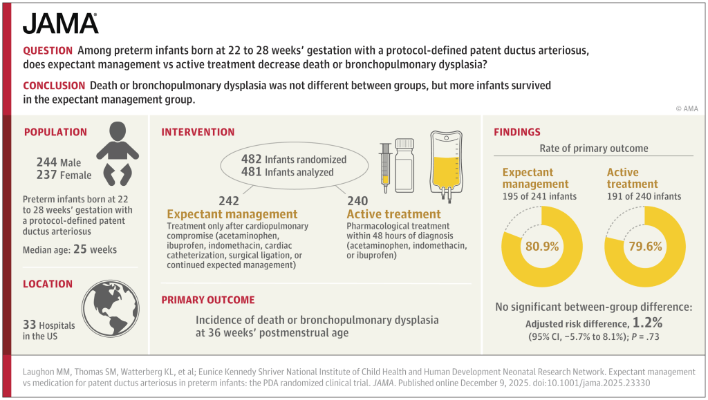
The breakdown of the primary outcome shows a lower mortality with expectant management than with intervention. At least, that is, mortality up to 36 weeks.
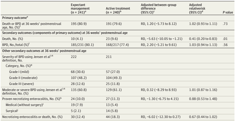
Surely the most important single question about any trial in sick preterm infants is : “If the baby receives treatment for the PDA, compared to expectant management, are they more likely to go home alive, or not?”

It takes a search of the supplemental materials, supplemental document 3, eTable 9, to find a partial answer to that question. The answer is that, by discharge or transfer, or 120 days after randomization (there were 18 infants still in the NICU at 120 days, and the investigators terminated the data collection), there were 14 deaths of the 241 expectant treatment infants. In the active treatment group there were 26 deaths among the 235 who actually got active treatment, there were 5 babies in this group who were not treated within 48 hours, and we don’t know if they survived or not.

If I do an ad hoc ITT chi-square, removing the unreported infants who had data collection truncated at 120 days, (as we don’t know if they lived or died) then the mortality is 14/237 vs 26/234. Or 5.9% vs 11.1%, a risk difference of 5.2% (95% CI of -0.2% to 11%), in other words not “statistically significant”.

As regular readers will know, I don’t think the arbitrary cutoff of p<0.05 is a good way to define what is real or not, which is why I almost always put “statistically significant” in quotation marks. But still, surely it was important to know that there were 7 pre-discharge deaths, at least, of the 40 total deaths, that occurred after 36 weeks. And there may have been more deaths after 120 days.

This has been a recurrent problem in similar studies, in BabyOscar for example, (Gupta et al 2024 in the figures below) mortality was reported at 36 weeks, and I can’t find any data anywhere about survival to discharge.

This is vitally important, the most recent meta-analyses show that medical treatment of the PDA increases BPD. Infants with severe BPD, still ventilated at 36 weeks have a measurable late mortality. Surely that is an outcome that we should know about?

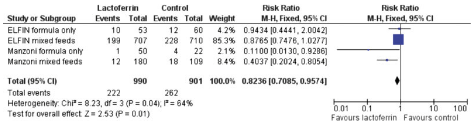
A recent SR/MA, published in May this year : Buvaneswarran S, et al. Active T “reatment vs Expectant Management of Patent Ductus Arteriosus in Preterm Infants: A Meta-Analysis. JAMA Pediatr. 179. United States2025. p. 877–85, included 10 trials of infants <33 weeks GA with “hemodynamically significant PDA (diagnosed by clinical or echocardiographic criteria)” who were randomized to active treatment compared to expectant management. There were a total of over 1000 babies per group and the primary outcome Forest plots are below
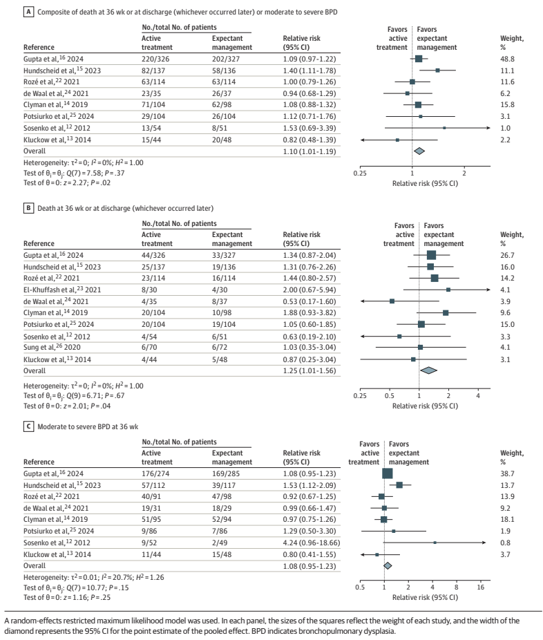
As you can see, this analysis suggests that there may well be an increase in “death at 36 weeks or at discharge (whichever occurred later)”, by which they actually mean whichever were the latest reported mortality figures.

They also include in the secondary outcomes, mortality before discharge, reported for less than half of the babies included in the various studies, which also shows an increase, but less marked, and with 95% CI including no difference.

To return to the new trial publication. I should make it clear that the trial was started in 2018, therefore probably designed in 2016 or so, and the authors are the brilliant Matt Laughon and the NICHD NRN centre representatives. The outcomes that they chose back then were the typical outcomes of PDA trials. It is easy for me, tapping away for my blog, to criticize, in retrospect, decisions that were taken a decade ago…

Nevertheless this trial, if you add it to the meta-analysis above, would surely confirm that closing a “haemodynamically significant” PDA does not appear to have any measurable benefits, and may well increase both mortality and oxygen requirements at 36 weeks, with most of the increase appearing to be in milder BPD, but probably a small increase in more severe disease also.

I am struggling to think of an evidence-based indication to close the PDA in a preterm infant. The data are, unfortunately rather muddy, there have been many protocol violations in the majority of the large trials. The exception was Hundscheid et al in the figure above, the BeNeDuctus trial, which only had 1 protocol violation in the expectant group. In BabyOscar, in contrast, 30% of the placebo group received open label medical or surgical PDA closure, about 1/3 of whom did not satisfy the protocol defined criteria. In the Rozé trial, 62% of the placebo babies had open label treatment, it is not clear how many satisfied their criteria for open-label treatment.

Which brings me to the question in the title, “Is there any indication for PDA closure?” The evidence-based answer to that question is that there are no criteria for defining a clinically important PDA, for which medical or surgical intervention has shown a survival benefit, or a reduction in lung injury.

I put it that way because there are some experts who continue to suggest that the big problem is with how we define a clinically important or “haemodynamically significant” PDA. (Bischoff AR, et al. Beyond diameter: redefining echocardiography criteria in trials of early PDA therapy. J Perinatol. 2025). And that all we have to do is better define the phenomenon. Which may be true, but requires that we prove it.

That recent opinion piece suggested that the criteria that should be used are those being tested in a pilot trial, the “Smart PDA” trial, which are not enormously different to the criteria used in the NICHD trial above. The authors of that piece remark that simply defining a significant PDA by diameter is insufficient; some PDAs with a large diameter have relatively modest impacts, and others of the same size may be associated with major shunts. The big difference between the Smart PDA trial, and the newly published NICHD RCT is that, in the newly published trial, being on CPAP, and having a PDA of over 1.5 mm would qualify for enrolment, without any other signs of a large shunt. In Smart PDA, you will also need at least one of the following signs of a L-R shunt

  1. Left atrium: aortic root ratio 1.5–2.0
  2. Transductal peak systolic velocity 1.5–2.0 m/s
  3. Left ventricular output (ml/kg/min) 200–400
  4. Diastolic flow pattern in the descending aorta: Absent/ retrograde

It seems to me that this has to be the next stage in the process, we should stop treating PDAs that do not have signs, such as those, of a substantial L-R shunt, doing so seems to have no benefit, and may well increase both oxygen requirements at 36 weeks, and perhaps even mortality. I think we have now reached the point where medical or surgical closure before 36 weeks PMA should only be attempted, in the context of an RCT, in infants with signs of a large shunt. There is currently no proven benefit to early PDA closure, and only harms.

In my recent practice I have seen some babies, usually “older” infants still ventilator dependent near term, with large shunts from a PDA, who have improved rapidly after ductal closure. In an NICU with 100 extremely preterm babies a year, there were maybe 3 or 4 in the last 4 years. It may be that those infants would have benefited from earlier closure, but in the absence of clearly defined criteria, which have been shown to predict benefit in preterm infants with a PDA, we frequently hesitate before performing a procedure with known risks. We have also had babies who have had little change in their clinical status after late PDA closure.

There do seem to be some babies who, anecdotally at least, seem to benefit from closure of the PDA. Our challenge as a community is to identify them, and hopefully to be able to identify them early enough that we can improve their outcomes.
Posted in Neonatal Research | Tagged , , , , , | 3 Comments

Is this article trustworthy?

There are many ways that an article in the medical literature may not be worthy of trust, and whose results may be unreliable. I remember one of my own trials, LACUNA, which was a pilot RCT of lactoferrin supplementation to prevent late-onset sepsis (LOS). As a pilot. the primary outcome was feasibility of a larger trial, but the clinical outcomes that I reported included the incidence of culture-positive sepsis. I calculated that as planned, as the proportion of babies who had at least one episode, out of the total number included in each group.

When I looked at the data, I realized that if I presented the results as infections per 100 patient days, there was actually a statistically significant difference between groups! I must admit to being tempted to report that outcome, as if it had been the primary outcome of my trial. I think the trial would have had a greater impact than it actually did, and could potentially have led to widespread adoption of lactoferrin as prophylaxis against late-onset neonatal sepsis. I could have been famous!

I am glad I resisted the temptation, because I know that it is vitally important to follow the trial design, outcomes, and statistical analysis that are planned prior to the trial. Any new analysis, or redefinition of outcomes, which is decided after examining the data, is inherently, extremely unreliable.

If I had reported that outcome, probably few people would have realized that I had changed the definition after performing the trial. The trial was pre-registered, but few people look at the registration documents, or compare the reported outcomes to those which were pre-planned. That includes peer reviewers, and meta-analysers.

It tuns out, of course, that the lactoferrin preparation that we used almost certainly has no real impact on LOS, and if I had reported the other outcome, it would eventually have been proven to be a red herring.

I was stimulated to think about this issue, which reminded me of the LACUNA outcomes, by a post from the Health Nerd https://gidmk.substack.com/p/the-terrifying-problem-of-fraud in women’s health. It is a really interesting blog, in general, and this post is indeed terrifying. He refers to several Cochrane reviews, including 2 that have major neonatal implications. He starts by discussing an influential Cochrane review of vitamin D supplements in pregnancy, that, in previous versions, was very positive, and led to recommendations from various health groups to give such supplements. He notes that the new version of the review has removed 21 articles because of trustworthiness issues. Twenty-one! The review now shows no benefit, and, in fact, potential harms.

A review with major neonatal impacts is the Cochrane review of steroids prior to Caesarian Delivery at term. The previous version included 4 trials, with nearly 4000 babies, and showed some significant benefits. The new version has removed 3 of those trials, for being untrustworthy, and now only shows a possible reduction in NICU admission among the 900 babies included in the 1 remaining trial, but not in RDS, or respiratory support. That single included trial did not report hypoglycaemia, which other trials, in late preterm infants, have shown to be a major potential complication of steroids in that population. Based on this new Cochrane review, I think such use of steroids is highly questionable.

Another Cochrane review from the obstetric group with a neonatal impact is their review of sildenafil and other NO pathway medications for fetal growth restriction. The initial search identified 19 studies, but 11 had to be excluded because of trustworthiness issues. Even one of those included was not prospectively registered, so they performed a sensitivity analysis excluding those data. Only sildenafil had enough data to be informative, and the review shows no benefit.

It is now essential that systematic review authors evaluate the trustworthiness of the trials being included. Almost all journals state that they follow the ICJME guidelines, which preclude the publication of trials which are not pre-registered. On many occasions however, I have seen articles published that are reports of clinical trials, but do not mention registration, or were retrospectively registered. Systematic reviews often include data from those trials without commenting on the issue. There should always be a sensitivity analysis excluding unregistered or retrospectively registered trials in a SR. The SR of erythopoietin for prevention of NEC, that I discuss in the post linked below, went from showing a reduction in NEC with erythropoietin, to a null effect when the retrospectively registered trials were deleted.

Just like my example above, untrustworthy research is not necessarily fraudulent. It may be a minor twist to the definition of the outcome, or deciding post hoc, that a certain observation is an outlier, or changing the analysis from treating a value as a dichotomous to a continuous outcome. Sometimes, of course, there is overt fraud, with entirely imaginary numbers. The pressure on individuals in some countries to produce published research is so enormous, that there is no consideration of the downstream harms that can be caused by such fraudulent results. Or by results which are “tweaked” to show a benefit.

The problem of untrustworthy published research is not new, but I fear that it will continue to grow, with articles now being generated entirely by AI. The least we can do is to demand that all published RCTs are publicly registered, in a database including projected sample size, eligibility criteria, and primary outcomes.

Those of us performing systematic reviews must now perform trustworthiness assessments, using one of the published tools. It substantially increases the workload involved, but will also improve the reliability of the results.

Posted in Neonatal Research | Tagged , , , | Leave a comment

Hope for HIE

I was struck this morning by a new article in Pediatric Research a “Family Perspectives” written by the founder of the organisation that has the same name as the title of this post, “Hope for HIE“, Betsy Pilon (Pilon B. Family reflections: what’s next for hypoxic-ischemic encephalopathy (HIE)—a patient advocacy perspective. Pediatr Res. 2025). It is an eloquent cri-de-coeur, demanding a greater voice for parents of infants with HIE:

“families affected by HIE remain sidelined in advocacy and institutional representation. We are routinely excluded in neonatology priority setting, where patient stakeholders may be represented, but HIE voices are not. The exclusion of HIE families leads to research agendas that don’t reflect our questions, timelines that don’t reflect our realities, and policies that fall short of what our children and families need.”

I think she has a point, many of our current parent partners are families of preterm infants, understandably, as they often spend weeks or months with us. HIE babies usually have shorter stays in the NICU, but the impacts on the families are just as great, and the long term impacts are sometimes greater. We should make extra efforts to ensure their voices are heard. She also points out some of the deficiencies of the longer term follow up of these children

“some families access follow-up to age two or three, very few have support as their children enter school, face academic challenges, or develop seizures, behavioral challenges, anxiety, or sensory processing issues. Research continues to overemphasize early developmental scores through assessments that are showing to not be predictive of neurocognitive development later in childhood when administered at age 2 or 3, and underemphasize the very issues that families identify as most critical in daily life”.

Any regular reader of this blog will know how much I agree with the limitations of early behavioural screening tests. Longer follow up of these infants is essential to both get a better picture of the impacts of HIE, but also to help the families to find the resources they need.

She ends with the following :

Max is now thirteen. He’s full of curiosity, humor, and resilience. He plays basketball, loves sushi, and is fiercely proud of how far he’s come. But he’s still living with the effects of HIE.
We all are.
Our journey didn’t end at discharge. It’s ongoing—and so is the work.
Let’s keep going, together.

Posted in Neonatal Research | Tagged , , | 1 Comment

Longer term outcomes after cooling

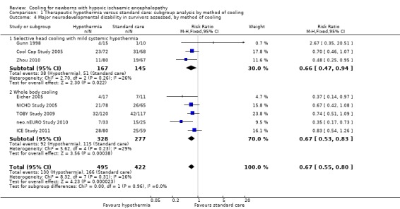
One of the numerous major advances in neonatology during my career has been the introduction of therapeutic hypothermia for infants with Hypoxic Ischemic Encephalopathy (HIE). Mortality is decreased, by about 25%, and long term morbidity among survivors is also decreased, by about 33%. Those estimates of effect size come from the Cochrane review, which provides the following Forest plot (I’m sorry about the quality of the image, the version in the pdf of the review is much clearer, but it extends over 2 pages, with a page break in the middle).

The Cochrane review also analyzed the impacts of cooling after dividing the infants according to severity of HIE, confirming that moderate and severe HIE both benefit. Unfortunately, the long term outcomes have been reported mostly up to 2 to 3 years. In the Cochrane review, 6 year outcomes are only available for the NICHD trial, which reviewed 120 survivors at 6 years, CP, IQ <70, executive function score <70, and moderate/severe disability were all lower in the hypothermia group than the controls, but the differences were small (and not “statistically significant”). The Cochrane review dates from 2013, and in 2014 the TOBY trial from the UK published follow up to 6-7 y of age, they showed more babies surviving without disability in the hypothermia group and “Among survivors, children in the hypothermia group, as compared with those in the control group, had significant reductions in the risk of cerebral palsy (21% vs. 36%, P=0.03) and the risk of moderate or severe disability (22% vs. 37%, P=0.03)”. Executive function scores were also higher in the cooled babies, and full scale IQ was 5 points higher (NS).

This new study has examined survivors of HIE and cooling sequentially, at 2y, 5y and 8 to 10 years, from 2 Dutch centres Parmentier CEJ, et al. Serial Assessment of Neurodevelopmental Outcome Following Neonatal Encephalopathy and Therapeutic Hypothermia. J Pediatr. 2025;285:114679. They used, appropriately, different tools at each age, each of which is normalised for the general population at that age, BSID ver 3, WPPSI, WISC, and other motor scales were used. The Child Behavior Checklist was also used for all children at the 2 later visits.

Scores on motor function scales were progressively worse as the children aged. Behavioural problems became more prevalent. Cognitive scores were overall fairly stable, but there was a progressive decrease in cognitive scores in the subgroup of infants who had damage to the mammillary bodies on MRI.

Another very recent article along the same lines, from Coimbra in Portugal, Vicente IN, et al. Neurodevelopment in the transition to school in children subjected to hypothermia due to neonatal hypoxic-ischemic encephalopathy: A prospective study. Early Hum Dev. 2025;207:106305) re-evaluated 39 survivors of cooling, who had been seen at 18-36 months, again at 48 to 78 months of age. They showed a shift to worse outcomes in the older assessments.

The changes were all due to deterioration in cognitive scores : “no further CP, epilepsy, or ASD diagnoses were made, cognitive performance declined in 11 children (28.0 %; p = 0.002; Wilcoxon test)”.

These studies point out the importance of much longer follow up of these children. They are an interesting contrast to preterm infants, who, overall, tend to have improved scores on standardized tests over time. The data on behavioural issues from the Dutch study, particularly increased internalizing behaviours, was interesting to me, as I was not really aware of this as a problem after HIE, also, behavioural problems are really important to families, and they may also be amenable to interventions to improve them.

These data make me wonder about the 2 issues of mild asphyxia, and the late preterm infant. If the prevalence of adverse outcomes changes so much over time, it may be that our decisions about which babies to cool are being influenced by somewhat unreliable data. There is very little longer term outcome data from the RCTs of cooling, to 5 to 6 years and beyond, that we might be missing a measurable benefit of cooling in such subgroups.

It is vital that trials of cooling for HIE, in groups for whom it is not yet proven to be beneficial, continue to follow the participants at least until early school age, and preferably towards adolescence. Only then will we be able to develop reliable data on the risks and benefits.

Posted in Neonatal Research | Tagged , , | 2 Comments

Face masks for resuscitation?

Blank DA, et al. Face mask versus nasal mask device use for initial resuscitation in extremely and very preterm infants (FONDUE): an open-label, single-centre, randomised, controlled trial. Lancet Child Adolesc Health. 2025;9(10):715–23.

An excellent acronym for this trial. Hopefully it will lead to a trend in acronyms based on European culinary specialities. Very preterm infants, n=151, of 23 to 32 weeks GA were randomized to receive delivery room CPAP with a face mask, or with a nasal mask in a single centre study from Monash in Melbourne. Delayed clamping was attempted, without respiratory support, or immediate clamping if the baby needed intervention. If the baby needed positive pressure ventilation, that was delivered by face mask, the same in the two groups. When the babies could be placed on CPAP, they randomly had either a face mask placed, or a nasal mask.

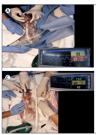
If the nasal CPAP was unsuccessful and the baby needed PPV, they were switched to a face mask. The authors supply some videos of the procedures, including this one of a baby started on nCPAP, then changed to face mask PPV.

The primary outcome of the trial was CPAP success defined as the “proportion of infants managed with CPAP only (ie, without positive pressure ventilation, intubation, chest compressions or adrenaline) between birth and transfer to the NICU. If a newborn received no respiratory support, that was considered success of the treatment group.” The proportion of CPAP successes are shown in the following table.

All the usual clinical outcomes were similar between groups. Admission FiO2 was lower in the nasal group.

It looks like the advantage of nasal, compared to face mask, CPAP was because more of the face mask group required PPV, 47/77, compared to 31/74 nasal mask subjects. This is consistent with previous findings that face mask application can cause apnoea. Stimulation of the trigeminal nerve area can provoke respiratory pauses, and bradycardia, and it seems that the nasal mask, creating pressure over a much smaller area around the base of the nose, does not have this effect. The authors note that the starting pressure was intended to be 5 to 8 cmH2O in the 2 groups, but that the clinicians started the nasal CPAP at an average of 1 cmH2O higher in the nasal group. The nasal group also had heated humidified gases, compared to the cold dry gases in the face-mask group. These 2 differences are potential confounding reasons for the difference between the 2 groups. But, because fewer nasal group babies had PPV, the peak inspiratory pressure applied was lower in that group than the face mask group.

Despite these limitations, it seems that there may well be significant advantages in applying a nasal mask, compared to a face mask, for CPAP in the delivery room in the extremely preterm infant. Although the authors did not show any improvement in clinical outcomes (the study was not powered for such outcomes), any intervention which decreases the need for PPV during transition is probably a good thing for lung protection.

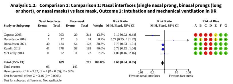
To put this in context of previous research, the Cochrane review, Ni Chathasaigh CM, et al. Nasal interfaces for neonatal resuscitation. Cochrane Database Syst Rev. 2023;10(10):CD009102, which did not include data from this trial, showed a reduction in the need for intubation in the DR with nasal interfaces compared to face mask. The 5 trials included in the Cochrane review were fairly heterogeneous: one included term infants; the nasal interface was a short nasal prong in 2 trials, short binasal prongs in 3 trials; and with a different device generating the pressure in the 2 groups in 2 of the trials. The Cochrane review showed a reduction in DR intubation, of note, this new trial had very few DR intubations, 6 in the nasal group and 7 in the face mask group; adding this to the Cochrane MA will have little impact, the weight will be small, and the tiny difference is in the same direction as the current MA results.

The review also showed less babies needing chest compressions, but that outcome was entirely dependent on the one trial that included full-term infants.

In the Donaldsson 2021 study the large majority of infants (<28 weeks) in both groups >82% received PPV. In Kamlin’s study, about the same proportion received non-intubated PPV (just over 50%) but fewer were intubated, McCarthy et al don’t seem to report how many infants needed PPV.

My interpretation of this is that it would be preferable in the very preterm infant to avoid face masks for initial CPAP support in the DR. It appears that the advantages of nasal prong systems and a nasal mask are similar, overall there is a reduction in the need for intubation in the DR, and perhaps for PPV.

Ni Chathasaigh CM, et al. Selective or routine face mask application for breathing support of preterm infants at birth: a randomised trial. Resuscitation. 2025;206:110467.

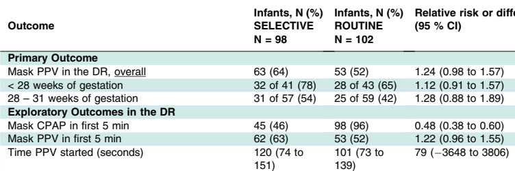
This RCT from Colm O’Donnell’s group at the national maternity centre in Dublin enrolled 200 babies <32 weeks. The idea was to determine if there was an advantage to routine immediate CPAP application, using a round Fisher-Paykell face mask, cold dry gases, and a t-piece resuscitator. The comparison, selective, group had face mask CPAP (using the same system) applied if they developed signs of respiratory distress after 5 minutes of age. Infants in both groups had standard resuscitation, with PPV being started if they were apnoeic, or if they had a heart rate <100. The primary outcome was the requirement for PPV. Although the difference in the primary outcome was small and not statistically significant, more babies in the selective group required PPV in the DR, in both GA strata, and nearly half of the selective group received early CPAP (before 5 minutes).

More extensive resuscitation (intubation, chest compressions) was very similar between groups, as were all the clinical outcomes after NICU admission. Although the study did not show any differences that were “statistically significant”, there were no benefits to delaying CPAP.

My interpretation of all this is that the very immature infant would probably most benefit from early CPAP applied with a nasal interface. I like the idea of using a nasal mask to avoid the potential trauma of inserting a nasal prong; prong insertion can usually be done gently, but sometimes, especially in the smallest babies it is a tight fit and probably hurts. A really useful trial would be to investigate routine early CPAP with a nasal mask, which can also be used for PPV, compared to using a face mask according to current NRP standards.

Posted in Neonatal Research | Tagged , , , , | 1 Comment

Beware articles written by AI

I subscribe to Google alerts, which sends me an email whenever the phrase “neonatal research” appears on a new website or a new post. I was interested, therefore to receive an alert about an article which, according the blog “Bioengineer.org”, showed a major genetic contribution to the occurrence of Necrotising Enterocolitis.

The blog post includes the following quote “Bai et al.’s study represents a landmark in neonatal research by providing compelling evidence for the heritability of necrotizing enterocolitis in very preterm infants. The twin study design elegantly disentangles genetic predisposition from environmental influences and firmly establishes a genetic foundation for this complex disease”.

This was intriguing, so I checked on the original article. (Bai R, et al. Genetic susceptibility to necrotizing enterocolitis in very preterm infants: evidence from twin data. Pediatr Res. 2025). A nice study, from a group of authors in China, one of whom is my good friend Shoo Lee, working with the Chinese Neonatal Network. They collected data on NEC incidence and chorionicity of twin pairs of less than 32 weeks GA (or <1500g). They found no difference in the likelihood of a coherent diagnosis of NEC between monochorionic and dichorionic twins. They did further analysis restricting to surgical NEC, or comparing early and late onset NEC, and found no difference between mono- and di-chorionic twins.

In other words, the actual findings of the study are exactly the opposite of what the post on that blog stated. The conclusion of the Bai et al authors was : “heritability does not play a major role in the development of NEC”.

I don’t think an actual human being, reading the article, could possibly have misinterpreted the findings quite as dramatically as whatever generated the blog post. The post is accompanied by the following cute image, which they note was AI generated. My only explanation for this dramatic misinterpretation of the original research article is that the post itself is also AI generated, and that the AI engine just loaded the title and some sub-headings from the results (which are, indeed, misleadingly worded as if there were positive findings : “Heritability contributes to NEC” and “Heritability contributes to certain subgroups of NEC”), without being able to realize that the actual results show that the analysis, of what should have been sub-titled “Heritability contribution to NEC”, was actually zero.

At least this is on an obscure blog, and will probably not cause any harm. In contrast, actual primary publications are also being generated by AI, reporting research that never actually happened. Government policy is also being influenced by review articles written by AI, which include non-existent research, or research which has been misinterpreted, often purposefully so, for partisan ends. This is a major issue for the future of medical research.

Let’s be careful out there.

Posted in Neonatal Research | Tagged | Leave a comment

Neonatal Research Shorts : November 2025

Ybarra M, et al. Low-Grade Germinal Matrix Hemorrhage-Intraventricular Hemorrhage and Concomitant Preterm Brain Injuries: Neurodevelopmental Outcomes at 3 Years of Age. J Pediatr. 2025:114713. Previous studies of the long term outcome of infants with germinal matrix or low grade IVH have been inconsistent. Some have shown an association with poorer developmental progress, and others have shown no impact. Some of this variability may be due to uncertainty about diagnostic criteria, with slightly larger amounts of intraventricular blood being classified differently. Some is probably due to the variable association with other brain injury, not readily seen on ultrasound, such as white matter injury, or cerebellar haemorrhages. We now routinely perform imaging of the posterior fossa, which was not easy with older ultrasound machines, but small cerebellar haemorrhages are still hard to see, without MRI.

In this cohort from Toronto, 175 infants <32 weeks GA had ultrasounds, they also had early cerebral MRI at 32 to 34 weeks, if they were stable, and then again at term equivalent age. Neurologic and developmental assessments were performed at 3 years (Bayley version III). As for the results, low grade haemorrhages had no correlation with outcomes, unless associated with either large cerebellar haemorrhages or more extensive white matter injury. It has always been fascinating to me that germinal matrix haemorrhage, which destroys the primary source of cortical neurones, has so little impact on long term outcomes. It speaks to the plasticity of the newborn brain, if the Germinal Matrix is injured, other parts of the brain take over neurone production.

Take home message : there was no apparent impact of GMH or small intraventricular haemorrhages without dilatation on long term development. Cerebellar haemorrhages, if large, are associated with delayed language development at 3 years, and white matter abnormalities, if extensive, are associated with motor delay, and cerebral palsy.

Price L, et al. Donor human milk pasteurization methods and the effect on milk components as they relate to Necrotizing enterocolitis. Semin Fetal Neonatal Med. 2025:101638.

Interesting review article on the impact of pasteurization, using the standard (Holder) pasteurization method, as well as some information about alternatives. The dash (-) in the figure above means no effect, rather than deletion. As you can see there are multiple impacts of pasteurization, as well as the expected impact on bacteria, some bacteria are resistant to Holder pasteurization, so donor breast milk still has an impact on the preterm intestinal microbiome. Both by direct colonization with the surviving organisms, but also because of the impact of HMOs and other components of human milk which remain despite pasteurization.

The figure also shows, in the upper right third, some alternative pasteurization methods which have been investigated, and which all show lesser impacts on breast milk components, HTST (high temperature short treatment) HPP (high pressure pasteurization) and UV-C (UV-C!). These alternative methods are equally effective at reducing bacterial load in the donor milk, and hopefully can be used in the future to give donor milk which is closer to Mothers Own Milk.

Take home message : Holder Pasteurization has major impacts on the composition of human milk. Alternative methods should be investigated, and approved.

Ottolini KM, et al. Donor human milk and structural brain development in very preterm infants. Pediatr Res. 2025.

In this observational study, the authors correlated the diet of a cohort of preterm infants <32 weeks GA with the findings on MRI at term. The cohort was enrolled over a long period, including a couple of years prior to the availability in their centre of donor milk (DHM) 2012-2014, and several years afterward 2014-2022. They include babies who almost exclusively received Mother’s own milk (MoM) and those receiving mostly formula, as well as the group with DHM. Brain volumes were greater in the human milk groups compared to formula, and diffusion tensor imaging showed diffusivity differences also, in the Corpus Callosum and the PLIC (posterior limb of the internal capsule). As the authors note, there is no good evidence from RCTs that DHM leads to better clinical neurological or developmental outcomes than formula. Nevertheless, these data are consistent with a beneficial effect of human milk on brain development, shared by DHM, and MoM.

Take home message : human milk seems to promote larger brains.

Wang H, et al. Bifidobacterium regulates premature infant gut metabolites, reducing serum inflammatory factors: a randomised controlled trial. Pediatr Res. 2025;97(3):1171–82.

One of the benefits of MoM is that it routinely contains probiotic organisms, usually including Bifidobacteria. In this trial, 70 preterm infants <32 weeks were randomized to control or to a supplement of Bifidobacterium animalis susp lactis. As often happens in some journals the article is written in somewhat strange English; one example : “Quality control and data analysis were conducted after instrument analysis, using assessment of the peak significante equation of standard curves”. They ran a statistical comparison of the baseline characteristics of the randomized groups. This is a practice that Pediatric Research should know is ridiculous. If the groups were randomized, why run such a statistical test? It is superfluous, potentially misleading, and the CONSORT statement specifically states that it should not be done, Pediatric Research is supposed to follow CONSORT guidelines.

I started to include this article in the post as I thought it was a demonstration of the possible anti-inflammatory impact of this Bifidobacterium on the preterm intestine. But I now realize that I haven’t got a clue what most of it means. This following figure for example, is supposed to show correlations between a large number of “metabolites”; about 30 were selected from over 250 that were found in the stools, including, for example, 34 different bile acids. These figures are supposed to show correlations, negative and positive, between “metabolites”.

The legend to the figure states “Red indicates positive correlation, blue indicates negative correlation, and the darker the colour, the stronger the correlation. a Probiotic group week 2 VS Control group week 2. b Probiotic group week 2 VS Control group week 2″…. What on earth is this supposed to mean? Aside from the fact that the potential of at least 62,500 comparisons were possible, is this comparison within the 2 groups, or between the 2 groups?

What it seems to show is that they measured a huge number of molecules, the concentrations of some of them were correlated with the concentrations of others. But so what?

They also show dramatic differences in serum TLR4 concentrations between groups. I am unsure if circulating concentrations of TLR4 are of any interest; TLR4 is normally attached to granulocytes, as part of the receptor complex which recognizes lipopolysaccharide. Nevertheless, published serum concentrations range between the pg/mL range to the mg/mL range, with these new results being intermediate in the ng/mL range. Such enormously variant normal ranges (over 1 million fold differences) make me very sceptical about any results. Serum TNF-α and IL-Iß were also dramatically lower in the Bifido group. They also give exactly the same data, regarding clinical complications, in table 3 and a completely superfluous figure 10.

Pediatric Research used to be a journal that only published high-quality research, although they were rarely clinical studies, which was previously one of my criticisms of the journal. If this is typical of the quality of what is currently getting through peer review and editorial control, then the journal has fallen far indeed.

Take home message : Pediatric Research is no longer the high-quality source it once was.

Kono Y, et al. Neurodevelopmental outcomes at age 3 years of preterm infants born at 22-31 weeks’ gestation. J Perinatol. 2025.

This is a report of the 3 year outcomes of babies from a cluster RCT of a quality control initiative in Japan. The original publication showed no impact of the QI program (INTACT), so the authors combined the groups for this publication describing their neurological and developmental outcomes. Babies were VLBW and ranged from 22 to 31 weeks GA. Below is a selection from the extensive results, “severely delayed” refers to being <70 on the cognitive subscale of the Kyoto Scales of Psychological Development. The KSPD seems to have a similar mean to the BSID ver3, when tested on the same infants, but have a wider distribution, so a score <70 was considered severely delayed.

There was very little blindness or deafness, so, as usual, it was cognitive delay which was responsible for most of the infants who were classified as “NDI”. Unfortunately, the authors don’t report many things which matter to parents, in particular there is no mention of behavioural problems. They do have a table that they call “functional outcomes” but that is actually a report of the medical interventions being received by the infants, at 3 years of age, including home oxygen, NG tube feeding, anticonvulsant medications, etc. All of which were rare.

Take home message : The majority of survivors at every gestational age, even the most immature, do not have “moderate or severe NDI”. There is a progressive increase in “moderate or severe NDI” as GA decreases.

Posted in Neonatal Research | Tagged , , , | Leave a comment

Clinical examination and treatment of septic shock. Not neonatology.

Andromeda-Shock-2 Investigators for the Andromeda Research Network SSoAR, et al. Personalized Hemodynamic Resuscitation Targeting Capillary Refill Time in Early Septic Shock: The ANDROMEDA-SHOCK-2 Randomized Clinical Trial. JAMA. 2025.

This is a very interesting trial evaluating the usefulness of clinical assessment of the circulation in adults with septic shock in a large international multicentre trial. Patients with suspected sepsis, who required norepinephrine after 1 litre fluid bolus, and had an elevated serum lactate, were randomized. A standardized method of measuring capillary refill time was agreed upon,

CRT was assessed by applying firm pressure to the ventral surface of the distal phalanx of a finger, using a glass microscope slide. The pressure was increased until the skin was blank, maintained for 10 seconds, and then released. The time required to return to the normal skin color was measured with a chronometer and a refill time longer than 3 seconds was defined as abnormal

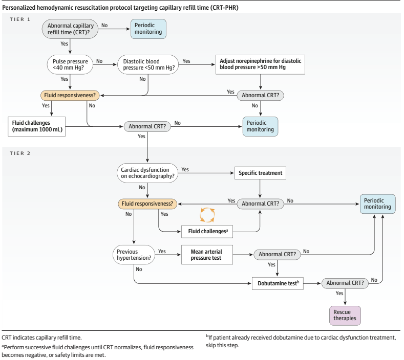
and the algorithms were activated if the cap filling time was abnormal in the CRT-PHR (cap refill time- personalized haemodynamic resuscitation) group.

As you can see, if the CRT was >3 seconds, you first check the pulse pressure, and if it is >40 mmHg, then you check the diastolic BP, which may lead to increasing norepinephrine dose; the next stage may be to give more fluid to see if there is a response, and then progress to bedside echocardiography, which may lead to specific treatments, or more fluid, or eventually to low dose dobutamine.

The control group had “standard care”, CRT was recorded but the algorithm was not followed.

The primary outcome was a hierarchical composite: (1) all-cause mortality within 28 days, (2) duration of vital support (vasoactives, mechanical ventilation, and kidney replacement therapy) truncated at day 28, and (3) length of hospital stay truncated at day 28.

The trial was analyzed by the Win Ratio. 1400 patients were randomized, as it was not a paired study (one way of using the Win Ratio), but individually randomized, they stratified the patients by APACHE score, then, within strata, every patient in group 1 was compared with every patient in group 2, to determine if they won or lost. There were therefore 244 000 paired comparisons. The CRT-PHR group won 49% of the comparisons, compared to 42% of the control, usual care group. The remaining 9% were exact ties.

This exceeded the limits for statistical significance; mortality was identical at 26.5%, but there were more ICU free days, and shorter hospital stays in the CRT group. The table of interventions shows that more of the CRT group received vasopressin, more received dobutamine, and they received less fluid; at 6 hours of treatment, their CRT was shorter, and serum lactate was lower.

The analysis is illustrated below in the 2 strata of the Apache Score (a higher score predicting higher mortality); this showed a greater difference in the sicker patients.

I found this fascinating. In terms of the intervention being investigated, trial design, and analysis methodology.

Many of my readers will know of my concerns about the way we analyse composite outcomes in neonatology. Comparing “death or BPD”, “death or NDI”, “death or hiccups”, between randomized groups, as if they were of equal importance, and as if we were always sure that they would change in the same direction with an intervention. This trial is one of a growing trend to using hierarchical composites, with death being given the greatest weight in the analysis, followed by other clinical outcomes in descending order of importance. Clearly an example to be followed in neonatology.

As for the intervention, I remember from my years as a staff in the PICU that one of the best predictors of a good outcome after major cardiac surgery is that the nurse tells you the infant’s toes are warm! It is a better predictor of survival than measuring cardiac output, for example. (Here is a recent example Cruz G, et al. Capillary Refill Time and Serum Lactate as Predictors of Mortality and Postoperative Extracorporeal Membrane Oxygenation Requirement in Congenital Heart Surgery. Children (Basel). 2023;10(5)).

I don’t think that capillary filling is quite as useful in preterm infants, kept in incubators, with their immature control of vascular tone, but there is still some correlation between cap filling and blood flow measured by doppler in the SVC. (Osborn DA, et al. Clinical detection of low upper body blood flow in very premature infants using blood pressure, capillary refill time, and central-peripheral temperature difference. Arch Dis Child Fetal Neonatal Ed. 2004;89(2):F168–73). As you can see below there is a lot of scatter, but a higher SVC flow is correlated with shorter CRT.

Combining such clinical signs with the direction of change in serum lactate (the absolute value doesn’t help much in the first couple of days as it is often high after birth), urine output (also not much use immediately after birth), level of activity etc, seems to me to be likely to be important in determining treatment in septic babies also. But we have very few good randomized trials of treatment approaches in septic newborns.

This trial gives us some pointers of how we could reasonably design such a trial, with a structured algorithm of interventions, including clinical pointers and targeted functional echocardiography in some patients, and how to design and analyse the primary outcome. We could develop a consensus algorithm (it couldn’t really be evidence-based) and test against usual care, with a hierarchical composite outcome including death and brain injury and duration of intensive care support, for example.

Posted in Neonatal Research | Tagged , , , , | Leave a comment

Lactoferrin supplementation does not prevent late-onset sepsis in the preterm… or is it more complicated that that?

Many of my readers will remember the impressive results of the high-quality study by Paolo Manzoni, Manzoni P, et al. Bovine lactoferrin supplementation for prevention of late-onset sepsis in very low-birth-weight neonates: a randomized trial. JAMA. 2009;302(13):1421–8) which showed that routine supplementation of preterm infants with bovine lactoferrin (bLF) dramatically reduced late-onset sepsis.

Many of us were quite excited with this finding, and launched our own studies, I performed a pilot in my NICU, hoping to use the data to get funding for a confirmatory trial (Barrington KJ, et al. The Lacuna Trial: a double-blind randomized controlled pilot trial of lactoferrin supplementation in the very preterm infant. J Perinatol. 2016;36(8):666–9), and at about the same time ELFIN was started in the UK. LIFT then took place in Australia/NZ, and, more recently, a Canadian version of LIFT was performed to increase study numbers and power.

Unfortunately all of the large confirmatory studies have been completely null, without a hint of a benefit. Including LIFT-Canada, which is in submission so I won’t go into any details, but I can say that we did not show a benefit of bLF.

There continue to be some trials which do seem to show an effect of bLF, including this very new trial (Plaza-Astasio V, et al. Preventing Sepsis in Preterm Infants with Bovine Lactoferrin: A Randomized Trial Exploring Immune and Antioxidant Effects. Nutrients. 2025;17(19)). Just over 100 VLBW infants were randomized to bLF supplementation or control, prior to 72 hours of age, and followed for LOS, as well as lab tests of antioxidant and immunologic effects. LOS was defined as “Laboratory confirmed sepsis” after 72 hours. The authors followed the NeoKisses definitions, which, as far as I can tell, include so-called “clinical sepsis” without a positive blood culture, but in the supplementary materials of this new study there are the same number of organisms listed as the episodes of sepsis, that is 11 in the bLF group and 21 in the placebo group. In other words they showed a reduction in culture-positive sepsis.

The authors note that their breast feeding rates were lower than some of the other large trials, at around 75% compared to over 90% in the large trials, and suggest this as a possible explanation for the difference of their results compared to the larger RCTs. That seems to me doubtful, if bLF was only effective in formula fed babies, then they could not have shown such a large decrease. Ochoa and her collaborators have published an IPD meta-analysis of the VLBW infants enrolled in their 2 trials (see below) which suggested that the impact of bLF was much greater among babies with low human milk intake (11% bLF, 21% controls). Although they do indeed show that, what is strange is that their analysis shows that LOS was much more frequent in babies with a high human milk intake, either with bLF (35%) or in their controls (39%), which is hard to understand. Another secondary analysis, of the data from ELFIN and the original Manzoni trial, showed similar reductions in LOS by bLF among breast-milk fed and formula fed, or mixed feeds babies. The reductions in LOS by bLF were very small and consistent with random variation in ELFIN. The interaction term was not significant, suggesting that the reduction in LOS was similar regardless of feed type.

The authors of the new study also note that their control frequency of sepsis was high, which is again true, a 40% incidence of LOS in a group of infants with a mean GA of 30 wks is extremely high. Having a higher baseline frequency of an abnormality will generally tend to make the impact of an intervention seem greater (see my recent posts on regression to the mean), but that doesn’t mean that such an impact would disappear completely when the incidence is lower.

One other difference that they do not mention is the source of bLF; the newly published trial used DicoPharm, just as did Manzoni. Akin’s study used the same product and also showed a reduction in culture-positive sepsis. Theresa Ochoa in her 2 studies used a product from Tatua ™ in the first study, derived from pasteurized milk, which had no effect on culture-positive sepsis, and a product from Friesland Campina in the other trial, which seems to be extracted by freeze-drying and not heat treated. The second trial showed a decrease in culture positive sepsis (from 11 to 8%, NS) not shared by the first study. Other studies either don’t mention the source of the bLF (Kaur et al) or I cannot obtain them as they aren’t in PubMed, or any other database that I can access (Liu, Tang, Dai). Another new study, from Egypt, randomized only formula fed infants (Ellakkany N, et al. Influence of bovine lactoferrin on feeding intolerance and intestinal permeability in preterm infants: a randomized controlled trial. Eur J Pediatr. 2024;184(1):30). They had an enormously high rate of LOS in the controls (60%) and a lower, but still extremely high, incidence in the bLF treated infants, 43.3%. The preparation they used was produced in Egypt, and I can’t find any details of how it was prepared. Finally I found one other trial, performed in Pakistan in infants with an average GA of about 34 weeks, (Ariff S, et al. Evaluation of Bovine Lactoferrin for Prevention of Late-Onset Sepsis in Low-Birth-Weight Infants: A Double-Blind Randomized Controlled Trial. Nutrients. 2025;17(11)). with a product from Hilmar in the USA which appears to have been prepared from freeze-dried milk (and perhaps not heat treated), they had an 8% incidence of culture-positive LOS in controls, and a combined 6% in the 2 treatment groups (with 2 different doses of bLF); total n of about 300.

There are lab studies showing that pasteurization decreases the biologic activity of bLF. bLF is degraded by heat treatment, it aggregates, and bind iron less well (Remadevi R, Mead D. A Study on the Bioavailability of Lactoferrin under Pasteurisation at Different Conductivities and Solid Contents. Journal of Food Research. 2025;14(2)). It could well be that heat-treatment of milk, prior to extraction of bLF, causes sufficient structural changes in the molecule for it to no longer have the multiple beneficial effects on bacterial proliferation that have been documented. This might be one reason why donor human milk (which is always pasteurized, usually by Holder pasteurization, the only method approved by HMBANA) is less effective at decreasing NEC than Mothers own Milk.

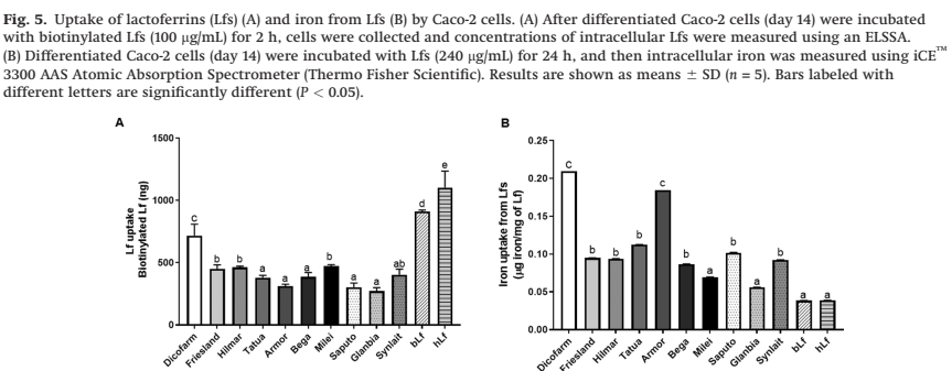
There are, however, known to be major differences in the biologic activity of different sources of bLF. One study examined 10 different bLF sources, and compared several different aspects of structure and activity between them, as well as their own bLF and human LF (Lonnerdal B, et al. Biological activities of commercial bovine lactoferrin sources. Biochem Cell Biol. 2021;99(1):35–46). There were major differences between bLF sources. As one example, they examined uptake of the LF by Caco-2 cells, and whether the LF transported iron into those cells

The details of what that means are not that important here (partly because my own understanding is limited, but also because it isn’t certain what this particular aspect has to do with their biologic effect of decreasing infections), but what this does show is that different sources of bLF are extremely different. They also found very variable degrees of contamination of the bLF product with other proteins, the Hilmar product, as one example “contains a relatively low concentration of Lf and relatively high concentrations of a-S1-casein, a-S2-casein, and J domain- containing protein”, whereas the Dicopharm product had lots of LF and relatively less of the other proteins.

I think, before we give up completely on bLF supplementation as a potential way to decrease LOS in the preterm, there is room for another study, investigating specifically the Dicopharm product, which has been consistently associated with decreases in culture-positive LOS. It may be that the story of bLF to prevent LOS still has a twist in the tale.

Posted in Neonatal Research | Tagged , , | 1 Comment

Return to the Regression to the Mean

After my post on regression to the mean, and its importance in studies of apnoea therapy, I was thinking of other examples. Some which have been most evident to me are those which impact on areas of medicine that I have researched myself. One example, from many years ago now, looked at the haemodynamic effects of dopamine in sick preterm infants. Seri I, et al. Regional hemodynamic effects of dopamine in the sick preterm neonate. J Pediatr. 1998;133(6):728–34.

This study was performed during the 1st 2 days of life, a period when blood pressure normally gradually increases, and when renal vascular resistance falls dramatically. These known baseline changes are an additional confounder in the results of non-controlled studies. The subjects were preterm infants with what they termed “compensated shock”, that is they had a BP between the 10th and 90th percentiles, but were oliguric (<0.6 ml/kg/h of urine) and/or had slow capillary filling. They were all given dopamine, with echographic indices performed before and after.

What you can see is that overall mean BP increased, after doses of dopamine between 2.5 and 7.5 microg/kg/min

And an index of renal vascular resistance, the pulsatility index in the renal artery, decreased

These are actually changes that you would expect over time in the first hours of life. The time difference between the 2 measurement was relatively short, at about 30 minutes, one could argue, perhaps, that the changes are too quick to just be postnatal adjustment. Maybe they were caused by the dopamine?

Interestingly, the authors also presented results after, post hoc, dividing the infants into responders (who had a >10% increase in mean BP) and non-responders.

This shows that, the “responders”, panel A, had a lower mean BP before dopamine treatment, of about 35, and it increased to about 43 afterwards. The “non-responders”, panel D, had a mean BP, before and after dopamine, of just over 40 mmHg.

This is exactly what you would see if the results are entirely due to regression to the mean. Those with lower BP than average will tend to have an increase after any treatment, including placebo. It would be surprising, in an observational study such as this, for them to have given dopamine to babies with a higher BP than average.

Having said that, dopamine will in some circumstances, I think it is clear, increase BP, probably not by much at a dose of 2.5, but there is enormous variability in dopamine kinetics (and pharmacodynamics); some infants might have an increase in BP at low doses, and some have no effect at very high doses. Dopamine is, however, an effective vasoconstrictor, and any increase in BP is entirely due to vasoconstrition in the newborn. In this study, however, both “responders” and “non-responders” had a decrease in renal vascular resistance, why would this be? As I mentioned above, renal vascular resistance is known to decrease dramatically after birth; this study, for example shows an 88% decrease in RVR over the first 2 weeks of life, most of which is in the first 2 to 3 days.

Additionally, there is no newborn animal model in which dopamine causes renal vasodilatation; indeed the “dopaminergic effect” on renal blood flow has only really been shown in healthy adult dogs! In newborn mammals, in contrast, only renal vasoconstriction has been shown with dopamine. Here is one of my studies, from many years ago (Pearson RJ, et al. Dopaminergic receptor-mediated effects in the mesenteric vasculature and renal vasculature of the chronically instrumented newborn piglet. Crit Care Med. 1996;24(10):1706–12). The piglets I used were relatively insensitive to dopamine, requiring more than 16 microg/kg/min to have a BP increase, but there was no decrease in RVR at any dose.

I also subjected the animals to an infusion of Fenoldopam, as selective agonist of vascular dopamine receptors, which showed absolutely no renal vasodilatation.

These examples demonstrate, yet again, that one has to be very sceptical about the results of observational studies of the responses to an intervention. Whenever we treat a baby who has a problem which varies in intensity, be it apnoea, low blood pressure, oxygen requirements, oliguria, or anything else that you can think of, unless you randomize and treat only half of the infants, one can never know if any changes which are seen are due to the intervention, or just regression to the mean. Babies with BP lower than average will always tend to have higher BP the next time you measure it. Babies with low urine output will always tend to have higher urine output after an interval.

Controls, controls, controls. Preferably randomized controls. They are essential for determining the impacts, efficacy and safety of our interventions.

Posted in Neonatal Research | Tagged , , , | 1 Comment